Discontinuity on T1-weighted image
and fluid signal on T2-weighted images
Fluid in the subacromial space on T2
images
Loss of subacromial fat plane on T1
Proliferative spur formation of
acromion or AC joint
Treatment
Conservative Treatment for
tendinopathy or chronic, partial tears
Cryotherapy
Rest
NSAIDs
Physical therapy (minimum of 6
weeks)
Initial stage – mobility
Second stage – strength
Third stage – function
Corticosteroid injections
Indications for Surgical Management
Acute, full-thickness tears in
normal rotator cuff
New functional deficit with a known,
partial tear
Arthroscopic Repair
“Full” Open Repair
“Mini” Open Repar
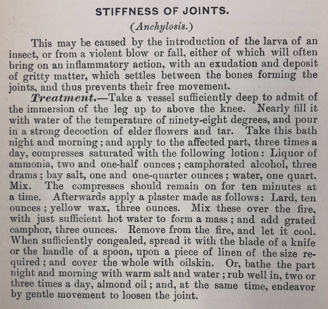
Cottage Physician
References
Oh LS, Wolf BR, Hall MP, Levy BA, Marx RG. Indications for rotator cuff repair: a systematic review. Clinical orthopaedics and related research. 2007; 455:52-63. [pubmed]
Teunis T, Lubberts B, Reilly BT, Ring D. A systematic review and pooled analysis of the prevalence of rotator cuff disease with increasing age. Journal of shoulder and elbow surgery. 2014; 23(12):1913-1921. [pubmed]
Lin TT, Lin CH, Chang CL, Chi CH, Chang ST, Sheu WH. The effect of diabetes, hyperlipidemia, and statins on the development of rotator cuff disease: a nationwide, 11-year, longitudinal, population-based follow-up study. The American journal of sports medicine. 2015; 43(9):2126-32. [pubmed]
Mehta S, Gimbel JA, Soslowsky LJ. Etiologic and pathogenetic factors for rotator cuff tendinopathy. Clinics in sports medicine. 2003; 22(4):791-812. [pubmed]
Sørensen AK, Bak K, Krarup AL, et al. Acute rotator cuff tear: do we miss the early diagnosis? A prospective study showing a high incidence of rotator cuff tears after shoulder trauma. Journal of shoulder and elbow surgery. ; 16(2):174-80. [pubmed]
Kim HM, Teefey SA, Zelig A, Galatz LM, Keener JD, Yamaguchi K. Shoulder strength in asymptomatic individuals with intact compared with torn rotator cuffs. The Journal of bone and joint surgery. American volume. 2009; 91(2):289-96. [pubmed]
Lew HL, Chen CP, Wang TG, Chew KT. Introduction to musculoskeletal diagnostic ultrasound: examination of the upper limb. American journal of physical medicine & rehabilitation. 2007; 86(4):310-21. [pubmed]
Roy JS, Braën C, Leblond J, et al. Diagnostic accuracy of ultrasonography, MRI and MR arthrography in the characterisation of rotator cuff disorders: a systematic review and meta-analysis. British journal of sports medicine. 2015; 49(20):1316-28. [pubmed]
Rees JD, Wilson AM, Wolman RL. Current concepts in the management of tendon disorders. Rheumatology (Oxford, England). 2006; 45(5):508-21. [pubmed]
Ainsworth R, Lewis JS. Exercise therapy for the conservative management of full thickness tears of the rotator cuff: a systematic review. British journal of sports medicine. 2007; 41(4):200-10. [pubmed]