
***LISTEN TO THE PODCAST HERE***
Aortic Stenosis

Causes
- Congenitally abnormal valve (bicuspid)
- Calcific disease –> most common in US
- Rheumatic valve disease –> most common world wide
Signs and Symptoms
- Dyspnea on exertion
- Exertional dizziness or syncope
- Angina
Description

- High-pitched, crescendo-decrescendo (diamond shaped), midsystolic murmur
- Soft S2
- S4 may be present
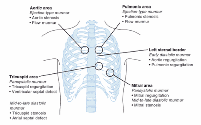
Best Auscultation Position
- Right 2nd intercostal space
Special Notes
- Radiate to carotid arteries
Aortic Regurgitation
Causes
- Aortic root dilation
- Congenital bicuspid valve
- Calcific disease
- Rheumatic heart disease –> most common world wide
Signs and Symptoms
- Exertional angina and yspnea
- Symptoms of heart failure
- PND, orthopnea, pulmonary edema, lower extremity edema
- Laterally and inferiorly displaced PMI with a thrill
Description

- Soft, high-pitched, early diastolic, decrescendo murmur
- Soft S1 with soft/absent A2
- S3 may be present
Best auscultation position
- Left 3rd intercostal space near sternal border (Erb’s point)
Special Notes
- Accentuated by patient sitting up and leaning forward at end expiration
Mitral Stenosis
Causes
- Rheumatic heart disease is most common causes
- Mitral annular calcification
- Radiation associated-valve disease (Hodgkin’s lymphoma)
Signs and Symptoms
- Exertional dyspnea
- Decreased exercise tolerance
- Hemoptysis (increased pulmonary pressure)
- Angina
- Fatigue
- Atrial fibrillation (elevated left atrial pressure)
- Hoarseness
Description

- Opening snap with low-pitched, diastolic murmur
- Decrescendo after S2
- Late, diastolic, crescendo before S1
- Loud S1
Best auscultation position
- Cardiac apex at left 5th intercostal space, midclavicular line
Special Notes
- Patient in left lateral decubitus in held expiration
- Using the bell
Mitral Regurgitation
Causes
- Primary
- Degenerative mitral valve disease à most common in US
- Mitral valve prolapse
- Rheumatic heart disease
- Infective endocarditis
- Trauma
- Congenital valve cleft
- Mitral annular calcification
- Degenerative mitral valve disease à most common in US
- Secondary
- Coronary artery disease (regional wall motion abnormality)
- Dilated cardiomyopathy
- Hypertrophic cardiomyopathy
Signs and Symptoms
- Exertional dyspnea
- Fatigue
- Atrial fibrillation
- Heart failure
Description

- High-pitched “blowing”, holosystolic murmur
- Diminished S1
Best auscultation position
- Cardiac apex at left 5th intercostal space, midclavicular line
Special Notes
- Radiates to axilla
- No variability in respiration
- Decreases in intensity with valsalva
Mitral Valve Prolapse
Causes
- Primary
- Sporadic (myxomatous degeneration)]\
- Familial (autosomal dominant with incomplete penetration)
- 30-50% in 1st degree relatives
- Secondary
- Connective tissue disorders
- 23CVInfective endocarditis
- Coronary artery disease
Signs and Symptoms
- Palpitations
- Dyspnea
- Exercise intolerance
- Dizziness or syncope
- Panic and anxiety disorders
- Numbness or tinging
Description

- Midsystolic click followed by a uniform, high-pitched, late systolic murmur
Best auscultation position
- Cardiac apex at left 5th intercostal space, midclavicular line
Special Notes
- Responds to dynamic auscultation
- Increased in sudden standing
- Decreased in sudden squatting
Tricupsid Stenosis
Causes
- Rheumatic heart disease
- Atrial myxoma
- Carcinoid syndrome
Signs and Symptoms
- Abdominal discomfort
- Hepatic congestion and heptomegaly
- Fluttering sensation in neck caused by jugular venous pulse
- JVD, ascites, peripheral edema
Description
- Soft, high-pitched, mid-diastolic
Best auscultation position
- 4th intercostal space on the sternal border
Special Notes
- Increased during inspiration, squatting, or leg raise
Tricuspid Regurgitation
Causes
- Functional
- Dilation of right atrium and ventricle with dilation of tricuspid annular leaflet
- Pulmonary hypertension, left-sided heart failure, left-to-right shunt
- Dilation of right atrium and ventricle with dilation of tricuspid annular leaflet
- Valvular
- Valve damage from pacemaker or ICD
- Infective endocarditis
- Rheumatic heart disease
- Ischemic heart disease
Signs and Symptoms
- Majority are symptomatic
- Right-sided heart failure
- Hepatomegaly, hepatic congestion, ascites, hepatic venous hum, JVD, edema
Description

- High-pitched, holosystolic murmur
Best auscultation position
- 4th intercostal space on the sternal border
Special Notes
- Radiates to right sternal border
- Increases with inspiration, leg raises, or squatting
Pulmonic Stenosis
Causes
- Congenital (10% of children with congenital heart disease)
- Tetralogy of Fallot
- Noonan Syndrome
- Bicuspid valves
- Calcification
Signs and Symptoms
- Exertional dyspnea
- Right heart failure
Description
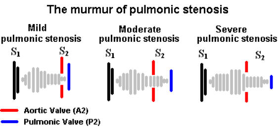
- Midsystolic, high-pitched, crescendo-decrescendo
- Pulmonary ejection click
- Extends through the A2
- Splitting of S2
Best auscultation position
- Left 2nd intercostal space
Special Notes
- Increased during inspiration
Pulmonic Regurgitation
Causes
- Primary
- Iatrogenic, infectious, rheumatic, congenital
- Secondary
- Pulmonary artery hypertension and/or dilation
- Physiologic (incidental)
Signs and Symptoms
- Asymptomatic until right ventricular dysfunction occurs
- Exertional dyspnea, fatigue
- Tachyarrythmias
Description
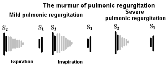
- Soft, high-pitched, early diastolic decrescendo
- Graham-Steele murmur (pulmonary HTN)
- High-pitched, blowing with accentuated P2
Best auscultation position
- Left 2nd intercostal space
Special Notes
- Increased with inspiration
S3 (ventricular gallop)
Causes
- Large amount of blood hitting a very compliant left ventricle
- Systolic heart failure
Description

- Low-pitched, early diastolic sound
- Occurs after S2
Best auscultation position
- Cardiac apex at left 5th intercostal space, midclavicular line
Special Notes
- Present with bell and absent with diaphragm
S4 (atrial gallop)
Causes
- Blood striking a non-compliant left ventricle
- Diastolic heart failure, LVH
Description
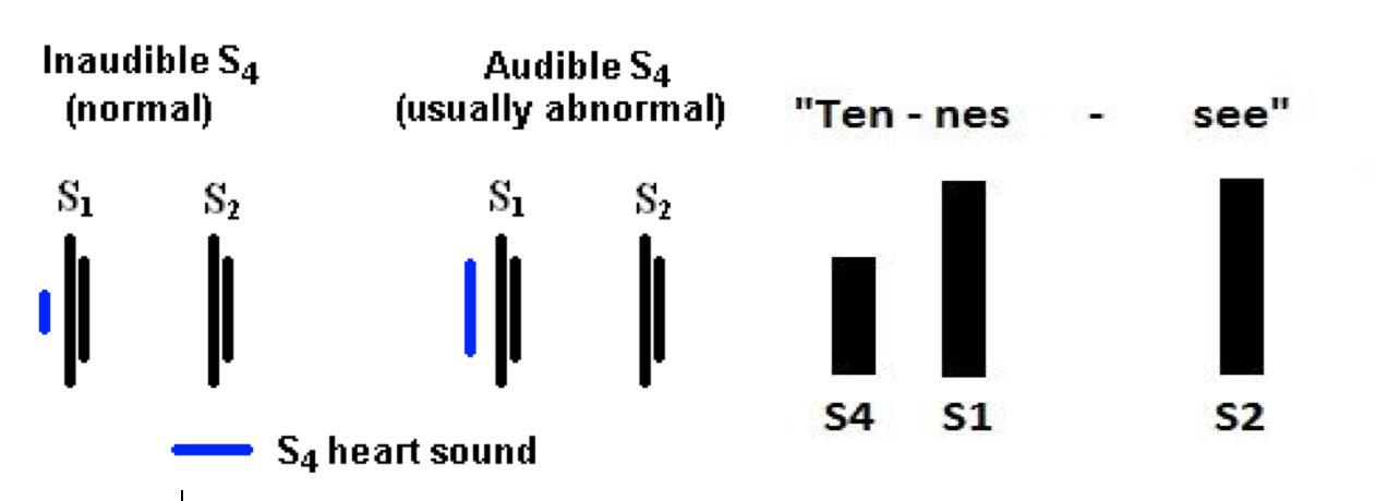
- Low-pitched, late-diastolic murmur
Best auscultation position
- Cardiac apex at left 5th intercostal space, midclavicular line
Special Notes
- Present with bell and absent with diaphragm

Cottage Physician
References
- Eveborn GW, Schirmer H, Heggelund G, Lunde P, Rasmussen K. The evolving epidemiology of valvular aortic stenosis. the Tromsø study. Heart (British Cardiac Society). 2013; 99(6):396-400. [pubmed]
- Enriquez-Sarano M, Tajik AJ. Clinical practice. Aortic regurgitation. NEJM. 2004; 351(15):1539-46. [pubmed]
- Horstkotte D, Niehues R, Strauer BE. Pathomorphological aspects, aetiology and natural history of acquired mitral valve stenosis. European heart journal. 1991; 12 Suppl B:55-60. [pubmed]
- Hull MC, Morris CG, Pepine CJ, Mendenhall NP. Valvular dysfunction and carotid, subclavian, and coronary artery disease in survivors of hodgkin lymphoma treated with radiation therapy. JAMA. 2003; 290(21):2831-7. [pubmed]
- Chandrashekhar Y, Westaby S, Narula J. Mitral stenosis. Lancet (London, England). 2009; 374(9697):1271-83. [pubmed]
- Freed LA, Benjamin EJ, Levy D. Mitral valve prolapse in the general population: the benign nature of echocardiographic features in the Framingham Heart Study. Journal of the American College of Cardiology. 2002; 40(7):1298-304. [pubmed]
- Strahan NV, Murphy EA, Fortuin NJ, Come PC, Humphries JO. Inheritance of the mitral valve prolapse syndrome. Discussion of a three-dimensional penetrance model. The American journal of medicine. 1983; 74(6):967-72. [pubmed]
- Sagie A, Schwammenthal E, Padial LR, Vazquez de Prada JA, Weyman AE, Levine RA. Determinants of functional tricuspid regurgitation in incomplete tricuspid valve closure: Doppler color flow study of 109 patients. Journal of the American College of Cardiology. 1994; 24(2):446-53. [pubmed]
- Snellen HA, Hartman H, Buis-Liem TN, Kole EH, Rohmer J. Pulmonic stenosis. Circulation. 1968; 38(1 Suppl):93-101. [pubmed]
- Nishimura RA, Otto CM, Bonow RO. 2014 AHA/ACC guideline for the management of patients with valvular heart disease: a report of the American College of Cardiology/American Heart Association Task Force on Practice Guidelines. Journal of the American College of Cardiology. 2014; 63(22):e57-185. [pubmed]
- Ozawa Y, Smith D, Craige E. Origin of the third heart sound. II. Studies in human subjects. Circulation. 1983; 67(2):399-404. [pubmed]
- Abrams J. Current concepts of the genesis of heart sounds. II. Third and fourth sounds. JAMA. 1978; 239(19):2029-30. [pubmed]
- Learn The Heart. https://www.healio.com/cardiology/learn-the-heart
