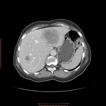
59yo male, with a history 27-pack-year history of smoking, presents to your primary care clinic for follow-up after a motor vehicle accident. He was evaluated in the local ED and was found to have a two masses on his liver on abdominal CT. What is the most likely cause of these lesions?
Answer
The most likely cause of this patient’s liver lesions is metastatic disease from distant primary malignancy. Hepatic metastases are 18-40 times more common than primary liver tumors due to the fact that liver is the primary filter for the body. In this particular patient, screening for lung cancer would be high on the list given his smoking history and a contrasted CT scan of the chest revealed a primary lung mass.
Ananthakrishnan A, Gogineni V, Saeian K. Epidemiology of primary and secondary liver cancers. Seminars in interventional radiology. 2006; 23(1):47-63. [pubmed]
Definition – mutated form of factor V that is unable to bind to protein C and leads to a hypercoaguable state
Clinical Significance – This is the most common hereditary hypercoaguability disorder in patients with European lineage. It increases the lifetime risk of DV, PTE, and stroke and patient are managed with lifelong anticoagulation.
History – Named after the Dutch city of Leiden where it was first discovered by Professor Rogier Bertina and Professor Pieter Reitsma in 1994 and subsequently published in Nature in their article entitled “Mutation in blood coagulation factor V associated with resistance to activated protein C”. Leiden has been one of Europe’s most prominent scientific centres for more than 400 years. It contains the oldest university in the Netherlands and has produced 13 Nobel Prize winners.
References
Firkin BG and Whitwirth JA. Dictionary of Medical Eponyms. 2nd ed. New York, NY; Parthenon Publishing Group. 1996.
Bartolucci S, Forbis P. Stedman’s Medical Eponyms. 2nd ed. Baltimore, MD; LWW. 2005.
Yee AJ, Pfiffner P. (2012). Medical Eponyms (Version 1.4.2) [Mobile Application Software]. Retrieved http://itunes.apple.com.
Bertina RM, Koeleman BP, Koster T, et al. Mutation in blood coagulation factor V associated with resistance to activated protein C. Nature. 1994; 369(6475):64-7. [pubmed]
59yo male, with a history 27-pack-year history of smoking, presents to your primary care clinic for follow-up after a motor vehicle accident. He was evaluated in the local ED and was found to have a two masses on his liver on abdominal CT. What is the most likely cause of these lesions?
Other Known Aliases – urine monoclonal globulin protein
Definition – immunoglobulin paraproteins produced by neoplastic plasma cells that are found in the urine due to decreased kidney filtration from acute kidney injury
Clinical Significance – Bence Jones proteins are classically associated with multiple myeloma and Waldenström’s macroglobulinemia and these proteins were detected by heating a urine specimen to promote precipitation of the protein, but now is seen on electrophoresis of concentrated urine. Newer serum free light chain assays have been shown to be more sensitive and superior to the urine studies and are coming into favor.
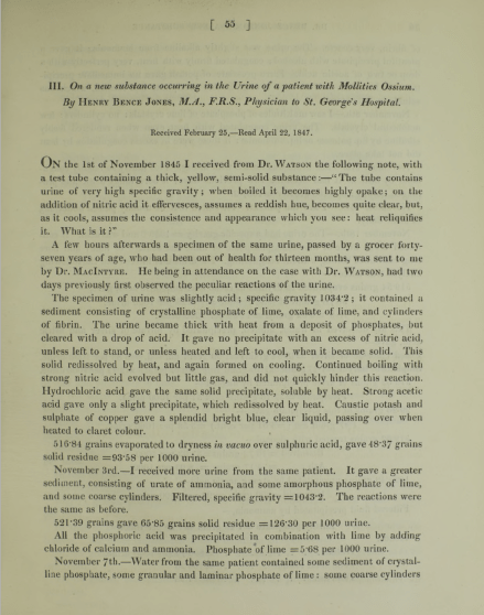
History – Named after Henry Bence Jones (1813-1873), who was an English physician and chemist and received his medical doctorate from St. George’s Hospital in 1840. His love for chemistry was sparked during his medical training and he simultaneously undertook private instruction in chemistry studies from professor Thomas Graham. After medical school, he went to Giessen, Germany to train under Justus von Liebig’s (the leading chemist of his time) at his animalistic chemistry school. He described his eponymous finding in 1847 in an article entitled “On a new substance occurring in the urine of a patient with Mollities Ossium”. His work on applying chemistry principles to human disease was so far ahead of his time that his work was not nearly as successful as it should have been due to the lack of knowledge of biochemistry and physiology of the time.
References
Firkin BG and Whitwirth JA. Dictionary of Medical Eponyms. 2nd ed. New York, NY; Parthenon Publishing Group. 1996.
Bartolucci S, Forbis P. Stedman’s Medical Eponyms. 2nd ed. Baltimore, MD; LWW. 2005.
Yee AJ, Pfiffner P. (2012). Medical Eponyms (Version 1.4.2) [Mobile Application Software]. Retrieved http://itunes.apple.com.
57yo Caucasian male presents to his primary provider with a one-year history of joint pain, weakness, and fatigue. He has a past medical history significant for hypertension and hyperlipidemia, for which he is being treated and is controlled on medications. His wife reports his skin has become a little darker over the last year as well. The rest of his physical examination does not reveal any abnormalities. Routine chemistries show a glucose of 214 mg/dL, AST of 472 mg/dL, and ALT of 513 mg/dL. What two (2) laboratory studies should be ordered next?
Answer
Hemochromatosis is at the top of the differential in a patient with elevated transaminases, heart disease, arthropathy, and hyperpigmented skin changes. As part of the initial screening, iron studies should be performed and an elevated transferrin saturation and serum ferritin are both elevated in hemochromotosis.
References
Bacon BR, Adams PC, Kowdley KV, Powell LW, Tavill AS, . Diagnosis and management of hemochromatosis: 2011 practice guideline by the American Association for the Study of Liver Diseases. Hepatology (Baltimore, Md.). 2011; 54(1):328-43. [pubmed]
Definition – pain in the posterior leg (classically behind the knee) with forced dorsiflexion of the foot
Clinical Significance – this examination finding was used in patients with a suspected DVT and before D-Dimers and clinical ultrasound were readily available. It is clinically useless as it has been studied extensively and found to have a sensitivity of 10-54% and specificity of 29-89%, thus not ruling in or out the condition consistently.
History – Named after John Homans (1877-1954), who was an American surgeon and received his medical doctorate from Harvard Medical School. He worked with Harvey Cushing and Samuel Crowe early in career exploring the connection between the piuitary gland and the reproductive system. He first described his eponymous finding in 1944 in a NEJM article entitled “Diseases of the veins” and later published the first case report of a DVT occuring after prolonged sitting on a flight between Boston and Caracas in 1954. He was a founding member of the the Society for Vascular Surgery and the namesake of the John Homans Chair of Surgery position at Harvard Medical School and John Homans Fellowship in Vascular Surgery at the Brigham and Women’s Hospital.
References
Firkin BG and Whitwirth JA. Dictionary of Medical Eponyms. 2nd ed. New York, NY; Parthenon Publishing Group. 1996.
Bartolucci S, Forbis P. Stedman’s Medical Eponyms. 2nd ed. Baltimore, MD; LWW. 2005.
Yee AJ, Pfiffner P. (2012). Medical Eponyms (Version 1.4.2) [Mobile Application Software]. Retrieved http://itunes.apple.com.
57yo Caucasian male presents to his primary provider with a one-year history of joint pain, weakness, and fatigue. He has a past medical history significant for hypertension and hyperlipidemia, for which he is being treated and is controlled on medications. His wife reports his skin has become a little darker over the last year as well. The rest of his physical examination does not reveal any abnormalities. Routine chemistries show a glucose of 214 mg/dL, AST of 472 mg/dL, and ALT of 513 mg/dL. What two (2) laboratory studies should be ordered next?
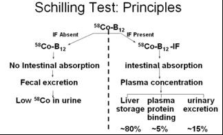
Definition – laboratory test for pernicious anemia (specifically intrinisic factor deficiency) that led to vitamin B12 (cobalamin) deficiency. It involved ingesting a oral dose of radiolabeled vitamin B12 (to test oral absorption), an IM injection of vitamin B12 (to saturate liver stores), and a 24hr urine collection to see how much was absorbed and excreted. If intestinal absorption was intact (intrinsic factor present), then > 10% of the radiolabeled vitamin B12 would be in the urine.
Clinical Significance – This was the first and only test at the time to be able to diagnose pernicious anemia, but is now largely a test of historical interest only as better diagnostic studies have been developed.
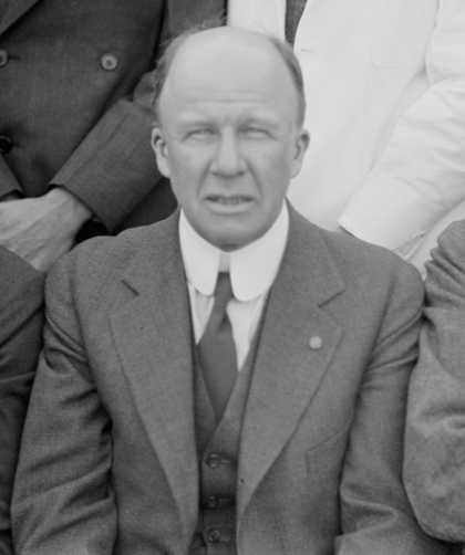
History – Named after Robert F. Schilling (1919-2014) an American physician and researcher who received his medical doctorate from the University of Wisconisn-Madison in 1943. Immediately after graduation, he joined the Pacific Front as a physician in the 3d Marine Division. After the war, he completed postgraduate training at Harvard before returning to Wisconsin to practice hematology. He studied extensively on the metabolism of vitamin B12 and the urinary excretion of radiolabeled vitamin B12 in pernicious anemia and in 1953, published a paper entitled “The effect of gastric juice on the urinary excretion of radioactivity after the oral administration of radioactive vitamin B12”, which would go on to be called the “Schilling Test”.
References
Firkin BG and Whitwirth JA. Dictionary of Medical Eponyms. 2nd ed. New York, NY; Parthenon Publishing Group. 1996.
Bartolucci S, Forbis P. Stedman’s Medical Eponyms. 2nd ed. Baltimore, MD; LWW. 2005.
Yee AJ, Pfiffner P. (2012). Medical Eponyms (Version 1.4.2) [Mobile Application Software]. Retrieved http://itunes.apple.com.
Schilling RF. Intrinsic factor studies. 2. The effect of gastric juice on the urinary excretion of radioactivity after the oral administration of radioactive vitamin B12. J Clin Lab Med. 1953;42;860-866