

***LISTEN TO THE PODCAST HERE***
Anatomy
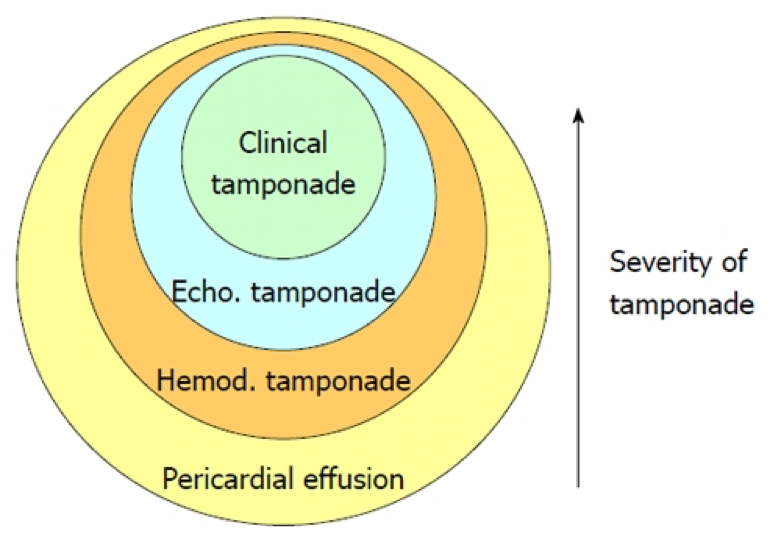
The pericardium consists of a double-layered semi-elastic sac that holds the heart in the mediastinum. Basically, so the heart doesn’t flop around inside the thoracic cavity. There should be a small amount of fluid (15-50mL) present to prevent adhesion of the pericardial sac to the heart. It is then termed an effusion when it is more than the normal amount. How much quantifies an effusion? Doesn’t matter…. what does matter is how fast that fluid develops. Because the pericardium is semi-elastic, it can accommodate and stretch over time if the accumulation is slow. This would lead to a greater volume of fluid before symptoms occur. If the fluid accumulates rapidly, less volume can produce profound effects due to the restrictive nature of the fibrous pericardium.

Etiology
- Infectious
- Viral
- Bacterial
- Fungal
- Parasitic
- Non-infectious
- Neoplastic
- Autoimmune/inflammatory
- Trauma
- Cardiac
- Radiation
- Metabolic
Signs and Symptoms
There are no reliable historical clues or physical exam findings that are specific to pericardial effusions. They are helpful, though, to sort out the cause of the effusion. Common findings include:
- Fever
- Dyspnea
- Chest pain
- Tachycardia
- JVD
- Hepatomegaly
- Pulsus paradoxus
-
- Ewart’s Sign
- Dullness to percussion, egophony, and bronchial breath sounds over the inferior angle of the left scapula
- Beck’s Triad
- Hypotension
- JVD
- Muffled heart tones

Work-Up
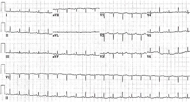
- EKG
- Sinus tachycardia
- Low voltage QRS (≤5mm in the 6 limb leads)
- Electrical alternans
-

Life In The Fastlane – o http://lifeinthefastlane.com/ecg-library/basics/low-qrs-voltage/
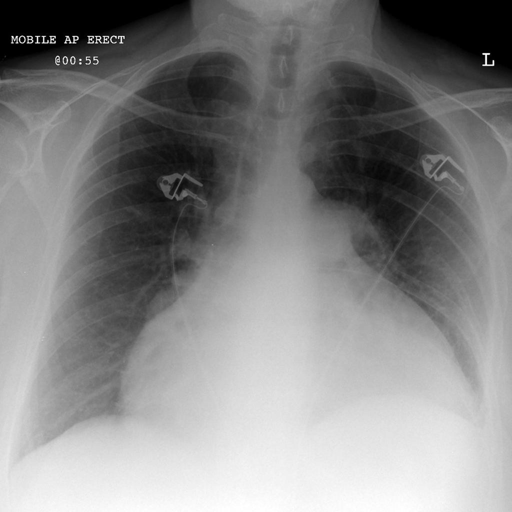
- Chest X-ray
- Small effusions are generally not appreciated on radiography
- Larger, chronic effusions may appear as an enlarged cardiac silhouette classically referred to as a “Water bottle heart”
-

Radiopaedia
- Echocardiogram
- Looking for anechoic stripe around the heart
- < 1cm ≈ 100cc
- 1-2cm ≈ 100-500cc
- > 2cm ≈ >500cc
- 2D Apical 4-chamber is my view of choice
- Severity can also be assessed by looking for:
- RV collapse during diastole
- LV collapse with increased EF
- IVC dilation and loss of respiratory variations
-
- Looking for anechoic stripe around the heart

Treatment
- Pericardiocentesis with catheter placement
-
- Three-way stopcock is used to measure pericardial pressure
- Fluid is then sequentially removed and pressure re-measured until < 5mmHg during inspiration
-
-
- Open surgical drainage via pericardial window if:
- Fluid accumulates after catheter drainage
- Effusion is loculated
- Need for biopsy
- Patient has coagulopathy
-
-
References
- Braunwald E. Pericardial Disease. In: Kasper D, Fauci A, Hauser S, Longo D, Jameson J, Loscalzo J. eds. Harrison’s Principles of Internal Medicine, 19e. New York, NY: McGraw-Hill; 2015. http://accessmedicine.mhmedical.com/content.aspx?bookid=1130&Sectionid=79743215. Accessed January 12, 2017.
- Imazio M. Contemporary management of pericardial diseases. Current Opinion in Cardiology. 2012;27(3):308-17. [pubmed]
- Levy PY, Corey R, Berger P. Etiologic diagnosis of 204 pericardial effusions. Medicine. 2003;82(6):385-91. [pubmed]
- Permanyer-Miralda G. Acute pericardial disease: approach to the aetiologic diagnosis. Heart (British Cardiac Society). 2004;90(3):252-4. [pubmed]
- Bruch C, Schmermund A, Dagres N. Changes in QRS voltage in cardiac tamponade and pericardial effusion: reversibility after pericardiocentesis and after anti-inflammatory drug treatment. Journal of the American College of Cardiology. 2001;38(1):219-26. [pubmed]
- Sternbach G. Claude Beck: cardiac compression triads. The Journal of Emergency Medicine. 1989;6(5):417-9. [pubmed]
- Stanford University. Tamponade. Echocardiography in ICU. https://web.stanford.edu/group/ccm_echocardio/cgi-bin/mediawiki/index.php/Tamponade.
- Adler Y, Charron P, Imazio M. 2015 ESC Guidelines for the diagnosis and management of pericardial diseases: The Task Force for the Diagnosis and Management of Pericardial Diseases of the European Society of Cardiology (ESC)Endorsed by: The European Association for Cardio-Thoracic Surgery (EACTS). European Heart Journal. 2015;36(42):2921-64. [pubmed]
- Gumrukcuoglu HA, Odabasi D, Akdag S, Ekim H. Management of Cardiac Tamponade: A Comperative Study between Echo-Guided Pericardiocentesis and Surgery-A Report of 100 Patients. Cardiology Research and Practice. 2011:197838. [pubmed]

Pingback: Today’s best #FOAMed #FOAMcc finds (2 – Jan 17) – Critical Care Northampton