Ep-PAINE-nym


Codman’s Triangle

Other Known Aliasesnone

Definitiontriangular area of new subperiosteal bone that is created when a bone tumor raises the periosteum away from the healthy bone
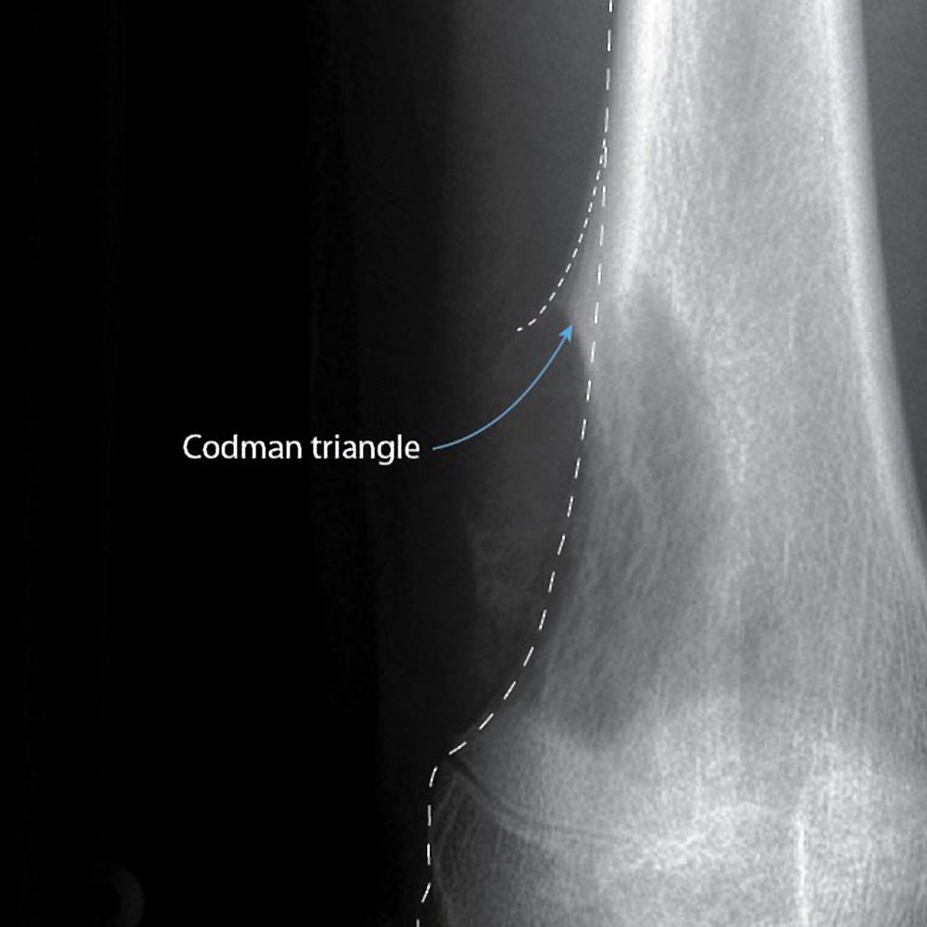
Clinical Significance this occurs because the tumor is growing at a faster rate than the periosteum can expand, which leads to the periosteum tearing away and providing a second edge of ossification (thus making the triangle). Presence of this finding is highly suggested of a fast growing, malignancy.

HistoryNames after Ernest Amory Codman (1869-1940), who was an American surgeon and received his medical doctorate from Harvard University in 1895. Aside from being an accomplished surgeon, he fought for hospital reform and was an early adopter and advocate for patient-based outcomes. In fact, he created “End Result Cards” for his patients which included all diagnosis, procedures, and treatment for every one of his patients that he tracked for at least one year. He was also the first physician at Massachusetts General Hospital to institute a morbidity and mortality conference. Unfortunately, he lost his surgical privileges when he wanted to institute a plan for evaluating surgical competence. He went on to found his own hospital based on end-results and published these findings to the general public in 1916. He established the first bone tumor registry in the US and helped lead the founding of the American College of Surgeons and its Hospital Standardized Program, which eventually became the Joint Commission on Accreditation of Healthcare Organizations.


References

  1. Firkin BG and Whitwirth JA.  Dictionary of Medical Eponyms. 2nd ed.  New York, NY; Parthenon Publishing Group. 1996.
  2. Bartolucci S, Forbis P.  Stedman’s Medical Eponyms.  2nd ed.  Baltimore, MD; LWW.  2005.
  3. Yee AJ, Pfiffner P. (2012).  Medical Eponyms (Version 1.4.2) [Mobile Application Software].  Retrieved http://itunes.apple.com.
  4. Whonamedit – dictionary of medical eponyms. http://www.whonamedit.com
  5. Up To Date. www.uptodate.com
  6. A Study in Hospital Efficiency. Boston : Privately printed, 1916.
  7. Bone Sarcoma, an Interpretation of the Nomenclature Used by the Committee of the Registry of Bone Sarcoma of the American College of Surgeons. New York : P. B. Hoeber, 1925.

PAINE #PANCE Pearl – Hematology


Question

You get a page from a gastroenterologist asking for a consult for platelet transfusion prior to an elective colonoscopy scheduled for tomorrow. The patient is a 56yo male, with a history of ITP and HTN. This procedure is for screening purposes only and the patient has not had any melena, hematochezia, or rectal bleeding. Platelet count is 97 x 109 per liter and the rest of the CBC is within normal limits.

  1. Does this patient meet the platelet transfusion threshold?
    1. If so, how much would you transfuse?
  2. If you were worried about bleeding, what are the other tests you could order to assess platelet function?

Answer

  1. The short answer is no, as he is not actively bleeding nor has any signs of anemia from chronic blood loss. Guidelines for platelet transfusions in preparation of invasive procedures is as follows:
    • < 100,000/microL for neurosurgery or ocular surgery
    • < 50,000/microL for most major surgery
    • < 50,000/microL for therapeutic endoscopic procedures
    • < 20,000/microL for low risk diagnostic endoscopic procedures
    • < 30,000/microL for bronchoscopy
    • < 20,000/microL for central line placement
    • < 20,000/microL for lumbar puncture with hematologic malignancy
    • < 50,000/microL for lumbar puncture without hematologic malignancy
    • < 80,000/microL for epidural anesthesia
    • < 20,000/microL for bone marrow biopsy
  2. The best way to assess platelet function is from a platelet function assay. This is generally determined by your facility’s lab, so it would be a good idea to talk with your lab to see what they use and how to interpret.


References

  1. Kumar A, Mhaskar R, Grossman BJ, et al. Platelet transfusion: a systematic review of the clinical evidence. Transfusion. 2015; 55(5):1116-27; quiz 1115. [pubmed]
  2. Warner MA, Woodrum D, Hanson A, Schroeder DR, Wilson G, Kor DJ. Preprocedural platelet transfusion for patients with thrombocytopenia undergoing interventional radiology procedures is not associated with reduced bleeding complications. Transfusion. 2017; 57(4):890-898. [pubmed]

Ep-PAINE-nym


Virchow’s Triad

Other Known Aliasesnone

Definitiontriad of broad categories of factors that contribute to thrombosis: hypercoaguability, endothelial injury, and stasis of blood flow

Clinical Significance These factors should always be considered in patients with suspected DVT, PTE, or acute arterial occlusion. Thought broad, they represent a simplistic mindmap to think of differential diagnoses and causes for patients with suspected conditions.

HistoryNames after Rudolf Ludwig Carl Virchow (1821-1902), who was a German physician and received his medical doctorate from the Friedrich-Wilhelms Institute in 1843. He had an interesting career in that he was a prolific writer (producing more than 2000 scientific manuscript), but also very politically charged and challenged not only the government, but also the status quo of medical education and dogmatism. This fervor allowed him to push the boundaries of what was known and being taught in medical schools and made him a well-known teacher, orator, and leader in the field of pathology. He first published his treatise on thrombosis in 1856 where he described his triad, but the eponym was not attributed to him until the mid-1900s.


References

  1. Firkin BG and Whitwirth JA.  Dictionary of Medical Eponyms. 2nd ed.  New York, NY; Parthenon Publishing Group. 1996.
  2. Bartolucci S, Forbis P.  Stedman’s Medical Eponyms.  2nd ed.  Baltimore, MD; LWW.  2005.
  3. Yee AJ, Pfiffner P. (2012).  Medical Eponyms (Version 1.4.2) [Mobile Application Software].  Retrieved http://itunes.apple.com.
  4. Whonamedit – dictionary of medical eponyms. http://www.whonamedit.com
  5. Up To Date. www.uptodate.com
  6. Virchow RLC. Gesammelte Abhandlungen zur wissenschaftlichen Medicin. Frankfurt am Main, 1 Meidinger, 1856+
  7. Bagot CN, Arya R. Virchow and his triad: a question of attribution. British journal of haematology. 2008; 143(2):180-90. [pubmed]

PAINE #PANCE Pearl – Hematology


Question

You get a page from a gastroenterologist asking for a consult for platelet transfusion prior to an elective colonoscopy scheduled for tomorrow. The patient is a 56yo male, with a history of ITP and HTN. This procedure is for screening purposes only and the patient has not had any melena, hematochezia, or rectal bleeding. Platelet count is 97 x 109 per liter and the rest of the CBC is within normal limits.

  1. Does this patient meet the platelet transfusion threshold?
    1. If so, how much would you transfuse?
  2. If you were worried about bleeding, what are the other tests you could order to assess platelet function?

Ep-PAINE-nym


Döhle Bodies

Other Known Aliasesnone

Definitionlight, blue-gray intra-cytosplasmic structures composed of agglutinated ribosomes most commonly found on neutrophils

Clinical Significance These inclusions are thought to be the remnants of the rough endoplasmic reticulum and represent defects in cell production and maturation during granulocytopoesis. As a result, Döhle bodies are seen in patients with infection, inflammation, and/or high physiologic stress, but may also be seen in pregnancy.

History – Named after Karl Gottfried Paul Döhle (1885-1928), who was a German pathologist and received his medical doctorate from the University of Kiel in 1882. He joined the faculty at his alma mater (where he would remain for his entire career) as an assistant to Arnold Ludwig Heller in 1883. He was an introvert by nature and rarely attended medical conferences and published very little of his work, but was well-renowned across his university. His work with Heller on describing syphilitic aortitis was groundbreaking and what eventually brought him contemporary fame in the field of histopathology. He published his findings on his eponymous cells in an article in 1892


References

  1. Firkin BG and Whitwirth JA.  Dictionary of Medical Eponyms. 2nd ed.  New York, NY; Parthenon Publishing Group. 1996.
  2. Bartolucci S, Forbis P.  Stedman’s Medical Eponyms.  2nd ed.  Baltimore, MD; LWW.  2005.
  3. Yee AJ, Pfiffner P. (2012).  Medical Eponyms (Version 1.4.2) [Mobile Application Software].  Retrieved http://itunes.apple.com.
  4. Whonamedit – dictionary of medical eponyms. http://www.whonamedit.com
  5. Up To Date. www.uptodate.com
  6. Döhle KGP. Vorläufige Mittheilung über Blutbefunde bei Masern. Zentralblatt für allgemeine Pathologie und pathologische Anatomie. Jena. 1892;3:150-152.

PAINE #PANCE Pearl – Oncology


Question

59yo male, with a history 27-pack-year history of smoking, presents to your primary care clinic for follow-up after a motor vehicle accident. He was evaluated in the local ED and was found to have a two masses on his liver on abdominal CT. What is the most likely cause of these lesions?

Answer

The most likely cause of this patient’s liver lesions is metastatic disease from distant primary malignancy. Hepatic metastases are 18-40 times more common than primary liver tumors due to the fact that liver is the primary filter for the body. In this particular patient, screening for lung cancer would be high on the list given his smoking history and a contrasted CT scan of the chest revealed a primary lung mass.


References

  1. Radiopaedia. Hepatic Metastases. https://radiopaedia.org/articles/hepatic-metastases-1
  2. Ananthakrishnan A, Gogineni V, Saeian K. Epidemiology of primary and secondary liver cancers. Seminars in interventional radiology. 2006; 23(1):47-63. [pubmed]

Ep-PAINE-nym


Factor V Leiden

Other Known Aliasesrs6025

Definitionmutated form of factor V that is unable to bind to protein C and leads to a hypercoaguable state

Clinical SignificanceThis is the most common hereditary hypercoaguability disorder in patients with European lineage. It increases the lifetime risk of DV, PTE, and stroke and patient are managed with lifelong anticoagulation.

HistoryNamed after the Dutch city of Leiden where it was first discovered by Professor Rogier Bertina and Professor Pieter Reitsma in 1994 and subsequently published in Nature in their article entitled “Mutation in blood coagulation factor V associated with resistance to activated protein C”. Leiden has been one of Europe’s most prominent scientific centres for more than 400 years. It contains the oldest university in the Netherlands and has produced 13 Nobel Prize winners.


References

  1. Firkin BG and Whitwirth JA.  Dictionary of Medical Eponyms. 2nd ed.  New York, NY; Parthenon Publishing Group. 1996.
  2. Bartolucci S, Forbis P.  Stedman’s Medical Eponyms.  2nd ed.  Baltimore, MD; LWW.  2005.
  3. Yee AJ, Pfiffner P. (2012).  Medical Eponyms (Version 1.4.2) [Mobile Application Software].  Retrieved http://itunes.apple.com.
  4. Whonamedit – dictionary of medical eponyms. http://www.whonamedit.com
  5. Up To Date. www.uptodate.com
  6. Bertina RM, Koeleman BP, Koster T, et al. Mutation in blood coagulation factor V associated with resistance to activated protein C. Nature. 1994; 369(6475):64-7. [pubmed]

Ep-PAINE-nym


Bence Jones Protein

Other Known Aliasesurine monoclonal globulin protein

Definitionimmunoglobulin paraproteins produced by neoplastic plasma cells that are found in the urine due to decreased kidney filtration from acute kidney injury

Clinical SignificanceBence Jones proteins are classically associated with multiple myeloma and Waldenström’s macroglobulinemia and these proteins were detected by heating a urine specimen to promote precipitation of the protein, but now is seen on electrophoresis of concentrated urine. Newer serum free light chain assays have been shown to be more sensitive and superior to the urine studies and are coming into favor.

HistoryNamed after Henry Bence Jones (1813-1873), who was an English physician and chemist and received his medical doctorate from St. George’s Hospital in 1840. His love for chemistry was sparked during his medical training and he simultaneously undertook private instruction in chemistry studies from professor Thomas Graham. After medical school, he went to Giessen, Germany to train under Justus von Liebig’s (the leading chemist of his time) at his animalistic chemistry school. He described his eponymous finding in 1847 in an article entitled “On a new substance occurring in the urine of a patient with Mollities Ossium”. His work on applying chemistry principles to human disease was so far ahead of his time that his work was not nearly as successful as it should have been due to the lack of knowledge of biochemistry and physiology of the time.


References

  1. Firkin BG and Whitwirth JA.  Dictionary of Medical Eponyms. 2nd ed.  New York, NY; Parthenon Publishing Group. 1996.
  2. Bartolucci S, Forbis P.  Stedman’s Medical Eponyms.  2nd ed.  Baltimore, MD; LWW.  2005.
  3. Yee AJ, Pfiffner P. (2012).  Medical Eponyms (Version 1.4.2) [Mobile Application Software].  Retrieved http://itunes.apple.com.
  4. Whonamedit – dictionary of medical eponyms. http://www.whonamedit.com
  5. Up To Date. www.uptodate.com
  6. Katzmann JA, Abraham RS, Dispenzieri A, Lust JA, Kyle RA. Diagnostic performance of quantitative kappa and lambda free light chain assays in clinical practice. Clinical chemistry. 2005; 51(5):878-81. [pubmed]
  7. Jones HB. On a new substance occurring in the urine of a patient with mollities ossium. Philosophical Transactions of the Royal Society. 1848;138:55–62. doi:10.1098/rstl.1848.0003

PAINE #PANCE Pearl – Hematology


Question

57yo Caucasian male presents to his primary provider with a one-year history of joint pain, weakness, and fatigue. He has a past medical history significant for hypertension and hyperlipidemia, for which he is being treated and is controlled on medications. His wife reports his skin has become a little darker over the last year as well. The rest of his physical examination does not reveal any abnormalities. Routine chemistries show a glucose of 214 mg/dL, AST of 472 mg/dL, and ALT of 513 mg/dL. What two (2) laboratory studies should be ordered next?


Answer

Hemochromatosis is at the top of the differential in a patient with elevated transaminases, heart disease, arthropathy, and hyperpigmented skin changes. As part of the initial screening, iron studies should be performed and an elevated transferrin saturation and serum ferritin are both elevated in hemochromotosis.


References

  • Bacon BR, Adams PC, Kowdley KV, Powell LW, Tavill AS, . Diagnosis and management of hemochromatosis: 2011 practice guideline by the American Association for the Study of Liver Diseases. Hepatology (Baltimore, Md.). 2011; 54(1):328-43. [pubmed]