Ep-PAINE-nym


Factor V Leiden

Other Known Aliasesrs6025

Definitionmutated form of factor V that is unable to bind to protein C and leads to a hypercoaguable state

Clinical SignificanceThis is the most common hereditary hypercoaguability disorder in patients with European lineage. It increases the lifetime risk of DV, PTE, and stroke and patient are managed with lifelong anticoagulation.
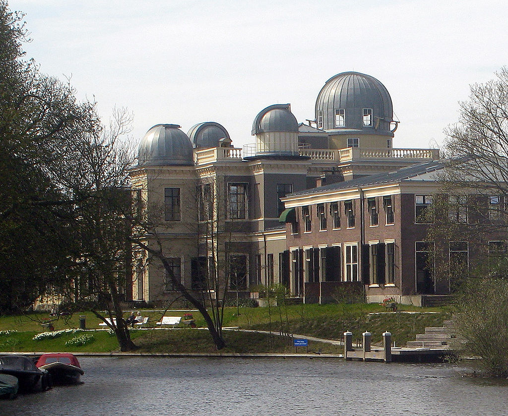
HistoryNamed after the Dutch city of Leiden where it was first discovered by Professor Rogier Bertina and Professor Pieter Reitsma in 1994 and subsequently published in Nature in their article entitled “Mutation in blood coagulation factor V associated with resistance to activated protein C”. Leiden has been one of Europe’s most prominent scientific centres for more than 400 years. It contains the oldest university in the Netherlands and has produced 13 Nobel Prize winners.


References

  1. Firkin BG and Whitwirth JA.  Dictionary of Medical Eponyms. 2nd ed.  New York, NY; Parthenon Publishing Group. 1996.
  2. Bartolucci S, Forbis P.  Stedman’s Medical Eponyms.  2nd ed.  Baltimore, MD; LWW.  2005.
  3. Yee AJ, Pfiffner P. (2012).  Medical Eponyms (Version 1.4.2) [Mobile Application Software].  Retrieved http://itunes.apple.com.
  4. Whonamedit – dictionary of medical eponyms. http://www.whonamedit.com
  5. Up To Date. www.uptodate.com
  6. Bertina RM, Koeleman BP, Koster T, et al. Mutation in blood coagulation factor V associated with resistance to activated protein C. Nature. 1994; 369(6475):64-7. [pubmed]

Ep-PAINE-nym


Bence Jones Protein

Other Known Aliasesurine monoclonal globulin protein

Definitionimmunoglobulin paraproteins produced by neoplastic plasma cells that are found in the urine due to decreased kidney filtration from acute kidney injury

Clinical SignificanceBence Jones proteins are classically associated with multiple myeloma and Waldenström’s macroglobulinemia and these proteins were detected by heating a urine specimen to promote precipitation of the protein, but now is seen on electrophoresis of concentrated urine. Newer serum free light chain assays have been shown to be more sensitive and superior to the urine studies and are coming into favor.

HistoryNamed after Henry Bence Jones (1813-1873), who was an English physician and chemist and received his medical doctorate from St. George’s Hospital in 1840. His love for chemistry was sparked during his medical training and he simultaneously undertook private instruction in chemistry studies from professor Thomas Graham. After medical school, he went to Giessen, Germany to train under Justus von Liebig’s (the leading chemist of his time) at his animalistic chemistry school. He described his eponymous finding in 1847 in an article entitled “On a new substance occurring in the urine of a patient with Mollities Ossium”. His work on applying chemistry principles to human disease was so far ahead of his time that his work was not nearly as successful as it should have been due to the lack of knowledge of biochemistry and physiology of the time.


References

  1. Firkin BG and Whitwirth JA.  Dictionary of Medical Eponyms. 2nd ed.  New York, NY; Parthenon Publishing Group. 1996.
  2. Bartolucci S, Forbis P.  Stedman’s Medical Eponyms.  2nd ed.  Baltimore, MD; LWW.  2005.
  3. Yee AJ, Pfiffner P. (2012).  Medical Eponyms (Version 1.4.2) [Mobile Application Software].  Retrieved http://itunes.apple.com.
  4. Whonamedit – dictionary of medical eponyms. http://www.whonamedit.com
  5. Up To Date. www.uptodate.com
  6. Katzmann JA, Abraham RS, Dispenzieri A, Lust JA, Kyle RA. Diagnostic performance of quantitative kappa and lambda free light chain assays in clinical practice. Clinical chemistry. 2005; 51(5):878-81. [pubmed]
  7. Jones HB. On a new substance occurring in the urine of a patient with mollities ossium. Philosophical Transactions of the Royal Society. 1848;138:55–62. doi:10.1098/rstl.1848.0003

PAINE #PANCE Pearl – Hematology


Question

57yo Caucasian male presents to his primary provider with a one-year history of joint pain, weakness, and fatigue. He has a past medical history significant for hypertension and hyperlipidemia, for which he is being treated and is controlled on medications. His wife reports his skin has become a little darker over the last year as well. The rest of his physical examination does not reveal any abnormalities. Routine chemistries show a glucose of 214 mg/dL, AST of 472 mg/dL, and ALT of 513 mg/dL. What two (2) laboratory studies should be ordered next?


Answer

Hemochromatosis is at the top of the differential in a patient with elevated transaminases, heart disease, arthropathy, and hyperpigmented skin changes. As part of the initial screening, iron studies should be performed and an elevated transferrin saturation and serum ferritin are both elevated in hemochromotosis.


References

  • Bacon BR, Adams PC, Kowdley KV, Powell LW, Tavill AS, . Diagnosis and management of hemochromatosis: 2011 practice guideline by the American Association for the Study of Liver Diseases. Hepatology (Baltimore, Md.). 2011; 54(1):328-43. [pubmed]

Ep-PAINE-nym


Homan’s Sign

Other Known Aliasesdorsiflexion sign

Definitionpain in the posterior leg (classically behind the knee) with forced dorsiflexion of the foot

Clinical Significancethis examination finding was used in patients with a suspected DVT and before D-Dimers and clinical ultrasound were readily available. It is clinically useless as it has been studied extensively and found to have a sensitivity of 10-54% and specificity of 29-89%, thus not ruling in or out the condition consistently.

HistoryNamed after John Homans (1877-1954), who was an American surgeon and received his medical doctorate from Harvard Medical School. He worked with Harvey Cushing and Samuel Crowe early in career exploring the connection between the piuitary gland and the reproductive system. He first described his eponymous finding in 1944 in a NEJM article entitled “Diseases of the veins” and later published the first case report of a DVT occuring after prolonged sitting on a flight between Boston and Caracas in 1954. He was a founding member of the the Society for Vascular Surgery and the namesake of the John Homans Chair of Surgery position at Harvard Medical School and John Homans Fellowship in Vascular Surgery at the Brigham and Women’s Hospital.


References

  1. Firkin BG and Whitwirth JA.  Dictionary of Medical Eponyms. 2nd ed.  New York, NY; Parthenon Publishing Group. 1996.
  2. Bartolucci S, Forbis P.  Stedman’s Medical Eponyms.  2nd ed.  Baltimore, MD; LWW.  2005.
  3. Yee AJ, Pfiffner P. (2012).  Medical Eponyms (Version 1.4.2) [Mobile Application Software].  Retrieved http://itunes.apple.com.
  4. Whonamedit – dictionary of medical eponyms. http://www.whonamedit.com
  5. Up To Date. www.uptodate.com
  6. McGee, Steven (2012). Evidence-Based Physical Diagnosis. Philadelphia, USA: Saunders. pp. 472–473. ISBN978-1-4377-2207-9
  7. Homans J. Diseases of the veins. N Engl J Med 1944: 231; 51-60
  8. Homans J. Thrombosis of the deep leg veins due to prolonged sitting. The New England journal of medicine. 1954; 250(4):148-9. [pubmed]
  9. Barker WF. John Homans, MD, 1877-1954. Arch Surg. 1999;134(9):1019–1020. doi:10.1001/archsurg.134.9.1019

PAINE #PANCE Pearl – Hematology


Question

57yo Caucasian male presents to his primary provider with a one-year history of joint pain, weakness, and fatigue. He has a past medical history significant for hypertension and hyperlipidemia, for which he is being treated and is controlled on medications. His wife reports his skin has become a little darker over the last year as well. The rest of his physical examination does not reveal any abnormalities. Routine chemistries show a glucose of 214 mg/dL, AST of 472 mg/dL, and ALT of 513 mg/dL. What two (2) laboratory studies should be ordered next?

Ep-PAINE-nym


Schilling Test

Other Known Aliases – none

Definitionlaboratory test for pernicious anemia (specifically intrinisic factor deficiency) that led to vitamin B12 (cobalamin) deficiency. It involved ingesting a oral dose of radiolabeled vitamin B12 (to test oral absorption), an IM injection of vitamin B12 (to saturate liver stores), and a 24hr urine collection to see how much was absorbed and excreted. If intestinal absorption was intact (intrinsic factor present), then > 10% of the radiolabeled vitamin B12 would be in the urine.

Clinical SignificanceThis was the first and only test at the time to be able to diagnose pernicious anemia, but is now largely a test of historical interest only as better diagnostic studies have been developed.

HistoryNamed after Robert F. Schilling (1919-2014) an American physician and researcher who received his medical doctorate from the University of Wisconisn-Madison in 1943. Immediately after graduation, he joined the Pacific Front as a physician in the 3d Marine Division. After the war, he completed postgraduate training at Harvard before returning to Wisconsin to practice hematology. He studied extensively on the metabolism of vitamin B12 and the urinary excretion of radiolabeled vitamin B12 in pernicious anemia and in 1953, published a paper entitled “The effect of gastric juice on the urinary excretion of radioactivity after the oral administration of radioactive vitamin B12”, which would go on to be called the “Schilling Test”.


References

  1. Firkin BG and Whitwirth JA.  Dictionary of Medical Eponyms. 2nd ed.  New York, NY; Parthenon Publishing Group. 1996.
  2. Bartolucci S, Forbis P.  Stedman’s Medical Eponyms.  2nd ed.  Baltimore, MD; LWW.  2005.
  3. Yee AJ, Pfiffner P. (2012).  Medical Eponyms (Version 1.4.2) [Mobile Application Software].  Retrieved http://itunes.apple.com.
  4. Whonamedit – dictionary of medical eponyms. http://www.whonamedit.com
  5. Up To Date. www.uptodate.com
  6. Schilling RF. Intrinsic factor studies. 2. The effect of gastric juice on the urinary excretion of radioactivity after the oral administration of radioactive vitamin B12. J Clin Lab Med. 1953;42;860-866
  7. Schilling Test. Stat Pearls. 2018. https://www.ncbi.nlm.nih.gov/books/NBK507784/

PAINE #PANCE Pearl – GYN


Question

Polycystic ovarian syndrome (PCOS) can often be a clinical diagnosis due to the classic distinguishing features of hirsutism, obesity, menstrual irregularities, and infertility. What is the classic relationship between FSH and LH in a patient with PCOS?


Answer

The classic relationship between LH and FSH in PCOS is > 2.5:1.

LH secretion is elevated, while FSH secretion is the same, or even decreased. LH stimulates theca cell proliferation and secretion of androgens, but there is insufficient FSH to stimulate granulosa cells. Although this is classically seen, LH:FSH is NOT used in any diagnostic criteria for PCOS.

Ep-PAINE-nym


Kruckenburg’s Tumor

Other Known Aliases – none

Definitionsecondary ovarian malignancy

Clinical SignificanceMost commonly arising from a gastric adenocarcinoma, but can occur from any metastatic cancer. 80% are bilateral and commonly manifest as pelvic pain, bloating, ascites, or dysparunea. Occasionaly, these tumor can be hormone producing and cause abnormal menstrual bleeding, hirsuitism, or virilization.

HistoryNamed after Friedrich Ernst Krukenberg (1871-1946), who was a German physician and received his medical doctorate from the University of Marburg.  He was actually studying to become a ophthalmologist, when he happend to be spending time in the pathology lab under Felix Marchand.  It was in this department that Krukenberg described a fibrosarcoma of the ovary (using sections from tumors Marchand had found in 1879) and published his findings in an article entitled “Über das Fibrosarcoma ovarii mucocellulare (carcinomatodes)” in 1896 at the age of 25 as part of his doctoral thesis. He spent the rest of career in his hometown of Halle, Germany practicing as a ophthalmologist.


References

  1. Firkin BG and Whitwirth JA.  Dictionary of Medical Eponyms. 2nd ed.  New York, NY; Parthenon Publishing Group. 1996.
  2. Bartolucci S, Forbis P.  Stedman’s Medical Eponyms.  2nd ed.  Baltimore, MD; LWW.  2005.
  3. Yee AJ, Pfiffner P. (2012).  Medical Eponyms (Version 1.4.2) [Mobile Application Software].  Retrieved http://itunes.apple.com.
  4. Whonamedit – dictionary of medical eponyms. http://www.whonamedit.com
  5. Up To Date. www.uptodate.com
  6. F. E. Krukenberg. Über das Fibrosarcoma ovarii mucocellulare (carcinomatodes).  Archiv für Gynäkologie. 1896;50:287-321.

#47 – Obstetrical Screening


*** LISTEN TO THE PODCAST HERE ***


Initial Prenatal Visit

  • Aneuploidy
    • American College of Obstetrics and Gynecology (ACOG) recommends:
      • All women should be offered screening before 20 weeks
      • All women should have the option for having a more invasive procedure instead of screening regardless of maternal age
        • Amniocentesis
        • Chorionic villus sampling
    • Two major categories of screening available
      • Specific maternal serum biomarkers
        • Primarily trisomy 21 and 18
      • Maternal circulation cell-free DNA
        • More sensitive
        • Assesses trisomy 21, 18, 13, and sex chromosome aneuploidies
  • Carrier Screening
    • ACOG recommends:
      • All women should be offered carrier screening for cystic fibrosis, spinal muscular dystrophy, thalassemias, and hemoglobinopathies
      • Fragile X
        • All women with a family history of intellectual disability, developmental delay, or autism
      • Each provider develop a screening strategies for ethnic-specific and panethnic populations
    • If there is a (+) screening test in the mother, then the reproductive partner should be offered screening
  • Standard Panel Laboratory Screening
    • ABO and Rh Screen
      • RhD(-) women should receive prophylactic anti(D)-immune globin at 28-weeks
    • Complete Blood Count and RBC Indices
      • 1st Screen for anemia
    • Documentation of Rubella and Varicella Immunity
      • Rubella IgG
      • Varicella IgG
    • Urinalysis and Urine Culture
      • Urine Protein – establish baseline to compare if patient develops pre-eclampsia or eclampsia
      • Untreated, asymptomatic has higher rates of developing pyelonephritis, pre-term birth
    • HIV Screen
      • ACOG recommends “opt-out” approach
    • Hepatitis B
      • HBsAg regardless of immunization status
    • Chlamydia
      • Nucelic Acid Amplification Test (NAAT) of endocervical/vaginal swab or urine
    • Syphilis
      • Can screen with either a non-treponemal or treponemal test, but a (+) screening test is confirmed with a treponemal test
  • Selective Screening in 1st Trimester
    • Thyroid Function – TSH only
    • Overt diabetes screening
      • Obtain HgbA1C if BMI > 25 (23 in Asian Americans) AND at least one of the following:
        • Gestational diabetes in previous pregnancy
        • HgbA1C > 5.7%, impaired glucose tolerance, or impaired fasting glucose on previous testing
        • 1st degree relative with diabetes
        • African-American, Latino, Native American, Asian American, Pacific Islander
        • History of cardiovascular disease
        • Hypertension (> 140/90 or on medication)
        • Age > 40yr
        • HDL cholesterol < 35 mg/dL or triglyceride > 250 mg/dL
        • PCOS
        • Physical inactivity
        • Other insulin resistance conditions
      • If HgbA1C > 6.5%, then treat as overt diabetes
      • If HgbA1C (-), then screen again at 24-28 weeks
    • Infections
      • Gonorrhea
        • NAAT from endocervical/vaginal swab
      • Hepatitis C
        • High risk patient should be screened with anti-HCV antibody or HCV RNA
      • Tuberculosis
        • Screen with tuberculin skin test or interferon-gamma release assay (IGRA) only if:
          • Suspicion for recent TB infection
          • Immunocompromised
      • Others
        • Toxoplasmosis, trichomonas, herpes simplex, cytomegalovirus, Zika, and Chagas are available for at risk patients or in endemic regions
    • Lead
      • Women with symptoms of lead exposure or risk factors

15-24 Weeks

  • These are not universal and are options available to mothers
  • Quadruple Test
    • Maternal serum alpha-fetoprotein level
    • Unconjugated estriol
    • Human chorionic gonadotropin
    • Inhibin A
  • Fetal ultrasound
    • Can be used to screen for neural tube defects and other fetal anomalies, as well as screen the mother for a short cervical length (< 25mm) that can increased her risk of spontaneous preterm birth

24-28 Weeks

  • Gestational Diabetes Screening
    • Two-Step Approach
      • Step One – Screening
        • 50g, one-hour glucose challenge test REGARDLESS of time of day or last meal
      • Step Two – Diagnostic
        • 100g, three-hour oral glucose tolerance test
          • Traditionally diagnostic after 2 elevated values, but newer data suggests that one may be OK
        • 75g, two-hour oral glucose tolerance test
          • Diagnostic after a single elevated value, but patient must be fasting
Up-To-Date
Up-To-Date
  • Complete Blood Count with iron and folate studies
    • 2nd anemia screening

28-36 Weeks

  • Sexually Transmitted Infection Screening
    • HIV, syphilis, chlamydia, gonorrhea, hepatitis B and C
    • Based on either previous (+) result or evidence of risk factors
Up-To-Date
  • Screen for group B beta-hemolytic streptococcus
    • Vaginal and rectal swabs
    • (+) results treated with intrapartum prophylaxis
CDC – GBS Prophylactic Antibiotic Algorithm
  • Screen for Fetal Growth Restrictions (<10th percentile weight for gestational age)
    • Indicated in third trimester in pregnancies at high risk
      • Infections, fetal anomalies, preeclampsia, gestational HTN and DM, alcohol use, placental/cord abnormalities

References

  1. ACOG Practice Bulletin No. 77: screening for fetal chromosomal abnormalities. Obstetrics and gynecology. 2007; 109(1):217-27. [pubmed]
  2. ACOG Practice Bulletin No. 88, December 2007. Invasive prenatal testing for aneuploidy. Obstetrics and gynecology. 2007; 110(6):1459-67. [pubmed]
  3. ACOG Committee Opinion No. 752: Prenatal and Perinatal Human Immunodeficiency Virus Testing. Obstetrics and gynecology. 2018; 132(3):e138-e142. [pubmed]
  4. Roberts SW, Sheffield JS, McIntire DD, Alexander JM. Urine screening for Chlamydia trachomatis during pregnancy. Obstetrics and gynecology. 2011; 117(4):883-5. [pubmed]
  5. Hughes BL, Page CM, Kuller JA. Hepatitis C in pregnancy: screening, treatment, and management. American journal of obstetrics and gynecology. 2017; 217(5):B2-B12. [pubmed]
  6. ACOG Practice Bulletin No. 190: Gestational Diabetes Mellitus. Obstetrics and gynecology. 2018; 131(2):e49-e64. [pubmed]
  7. Centers for Disease Control.  Group B Strep (GBS).  https://www.cdc.gov/groupbstrep/guidelines/new-differences.html
  8. Bricker L, Medley N, Pratt JJ. Routine ultrasound in late pregnancy (after 24 weeks’ gestation). The Cochrane database of systematic reviews. 2015; [pubmed]