47yo man presents to your clinic to establish care. He has a history of resistant hypertension, DMII, and sleep apnea. Vital signs are BP-159/101, HR-74, RR-16, O2-100%, and temp-98.9. Physical examination is also significant for multiple bruises on the lower extremities.
What would be the next step in the diagnosis of this patient?
What else would you need to order to determine the cause of this patient’s condition?
Definition – constellation of signs and symptoms due to excessive cortisol. This can be caused by several different mechanism that affect the hypothalamus-pituitary-adrenal axis:
CRH secretion by hypothalamus
ACTH secretion by:
Anterior pituitary
Ectopic tumor
Cortisol secretion adrenal glands by:
Adrenal hyperplasia
Adrenal tumor
Exogenous administration of corticosteroids
Clinical Significance – This is one of the more common endocrinologic pathologies you will see in clinical practice. Classic presentation includes obesity, abdominal striae, “moon face”, “buffalo hump”, and hirsutism. Diagnosis is made by obtaining a 24-hour urine cortisol measurement
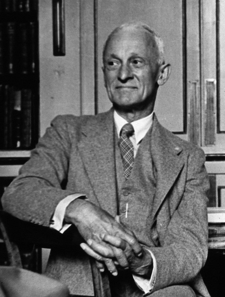
History – Named after Harvey Williams Cushing (1869-1939), who was an American surgeon and pioneering neurosurgeon of the early 20th century. He received his medical doctorate from Harvard Medical School in 1895. He completed his internship at Massachussets General Hospital and went on to do a surgical residency under William Halsted at John Hopkins Hospital. He trained under Kocher in England for several years before returning stateside and setting up practice in Baltimore. One of his greatest contributions to western medicine was his introduction of blood pressure management he learned from Scipione Riva-Rocci in Italy during his time in Europe.
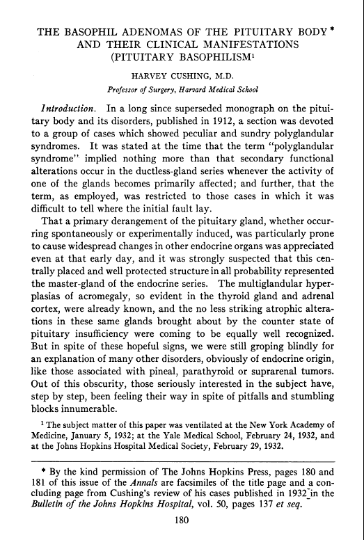
At the age of 32, he achieved associate professor at Johns Hopkins Hospital and was placed in full charge of all surgery of the central nervous system. In 1912, he first described what would become his eponymous disease, but before he could publish it, he was called to serve during the first world war as the director for a field hospital in France for the British. It was during this appointment that he cared for a fatally wounded soldier by the name of Lt. Edward Revere Osler, son of William Osler. He formally published his findings on his eponymous disease in 1932 in an article entitled “The Basophil Adenoma of the Pituitary Body and Their Clinical Manifestations: Pituitary Basophilism”.
During his career, he was regarded as the world’s leading teacher of neurosurgeons for in the first decades of the 20th century and held professorships at Johns Hopkins Hospital, Brigham Hospital in Boston, Harvard Medical School, Yale School of Medicine, as well as honorary Fellowship in the Royal College of Surgeons. He also was awarded the 1926 Pulitzer Prize for Biography for his biography on the life of William Osler and was nominated for the Nobel Prize in Physiology or Medicine 28 times.
Cushing (far left) with Osler (second from right) and Kelley (second from left). Johns Hopkins Hospital. 1900.
References
Firkin BG and Whitwirth JA. Dictionary of Medical Eponyms. 2nd ed. New York, NY; Parthenon Publishing Group. 1996.
Bartolucci S, Forbis P. Stedman’s Medical Eponyms. 2nd ed. Baltimore, MD; LWW. 2005.
Yee AJ, Pfiffner P. (2012). Medical Eponyms (Version 1.4.2) [Mobile Application Software]. Retrieved http://itunes.apple.com.
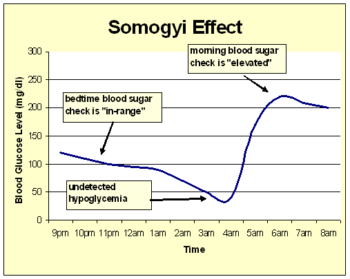
58yo
male, with DMI controlled with insulin, has blood glucose measurements
in the morning of 205-272 mg/dL for the past week. He reports that his
evening blood glucose measurements before bed range from 103-127 mg/dL.
What are two potential causes of these findings?
Answer
There are two potential causes of early morning hyperglycemia in a diabetic patient on insulin.
Dawn Phenomenon
Due to the early morning rise of cortisol, patients can experience an early morning hyperglycemia as a result
Somogyi Phenomenon
(as covered by last week’s Ep-PAINE-nym)
This was theorized to occur as undetectable hypoglycemia while the
patient was asleep and the resultant hyperglycemia from instrinsic
protective mechanisms.
Other Known Aliases – posthypoglycemic hyperglycemia
Definition – rebounding hyperglycemia in the setting of a undetected hypoglycemic event
Clinical Significance – It had been hypothesized that patients who had a hypoglycemic event during sleep would have rebound hyperglycemia due to the protective mechanism of the body to counteract this. This would result in an undetectable change that would cause hyperglycemia in the morning. This hypothesis has been proven wrong by numerous studies, but it is still a favorite among endocrinologists to pimp their students on.
History –Named after Michael Somogyi (1883-1971), who was a Hungarian American biochemist and received his doctorate degree from the University of Budapest in 1914. He took a position as professor of biochemistry in 1922 at the Washington University’s Medical School in St. Louis, where later that year the first child with diabetes was treated with an insulin prepared by Somogyi himself. His career work revolved around diabetes and theorized that insulin itself could causes unstable diabetes. He published this paper describing the phenomenon that bears his name in 1938 in the Weekly Bulletin of the St. Louis Medical Society entitled “Insulin as a cause of extreme hyperglycemia and instability”. He also went on to develop the test for serum amylase to help diagnose acute pancreatitis. Dr. Somogyi died from a stroke on July 21st, 1971.
References
Firkin BG and Whitwirth JA. Dictionary of Medical Eponyms. 2nd ed. New York, NY; Parthenon Publishing Group. 1996.
Bartolucci S, Forbis P. Stedman’s Medical Eponyms. 2nd ed. Baltimore, MD; LWW. 2005.
Yee AJ, Pfiffner P. (2012). Medical Eponyms (Version 1.4.2) [Mobile Application Software]. Retrieved http://itunes.apple.com.
M. Somogyi, “Insulin as a cause of extreme hyperglycemia and instability,” Weekly Bulletin of the St Louis Medical Society, 1938,
Tordjman KM, Havlin CE, Levandoski LA, White NH, Santiago JV, Cryer PE. Failure of nocturnal hypoglycemia to cause fasting hyperglycemia in patients with insulin-dependent diabetes mellitus. The New England journal of medicine. 1987; 317(25):1552-9. [pubmed]
Hirsch IB, Smith LJ, Havlin CE, Shah SD, Clutter WE, Cryer PE. Failure of nocturnal hypoglycemia to cause daytime hyperglycemia in patients with IDDM. Diabetes care. 1990; 13(2):133-42. [pubmed]
58yo male, with DMI controlled with insulin, has blood glucose measurements in the morning of 205-272 mg/dL for the past week. He reports that his evening blood glucose measurements before bed range from 103-127 mg/dL. What are two potential causes of these findings?
Definition – Hyperthyrodism caused by antibodies that stimulate T3/T4 secretion. The most common antibodies are thyroid-secreting hormone (TSH) and thyrotropin receptor antibody (TRAb).
Clinical Significance – Classic clinical manifestations of hyperthyroidism include thyromegaly, ophthalmaopathy, resting tremor, palpitations, weight loss, heat intolerance. For more in depth analysis of hyperthyroidism, see my 2017 talk at ASPA here.
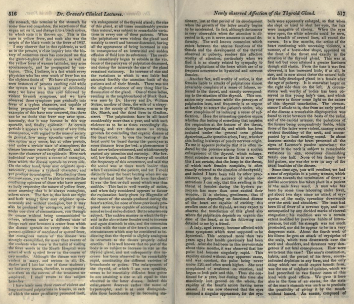
History –Named after Robert James Graves (1796-1853), who was an prolific Irish physician, surgeon, and educator. He was named Regius professor of the Institute of Medicine in Trinity College, founded the Dublin Journal of Medical and Chemical Sciences, and was a an early adopter of clinical bedside rounding and teaching with medical students. Dr. Graves wrote a routine clinical lecture series in the London Medical and Surgical Journal and first described a young female patient with ophthalmopathy and goiter in 1835. Dr. Armand Trousseau then published the collection of these articles in 1864 entitled “Clinical Lectures on the Practice of Medicine” and gave him this eponym. Another contribution of Dr. Graves was the creation of the second hand on watches to time pulses and the practice of providing food and water with patients with a fever, instead of the common practice of withholding nourishment.
References
Firkin BG and Whitwirth JA. Dictionary of Medical Eponyms. 2nd ed. New York, NY; Parthenon Publishing Group. 1996.
Bartolucci S, Forbis P. Stedman’s Medical Eponyms. 2nd ed. Baltimore, MD; LWW. 2005.
Yee AJ, Pfiffner P. (2012). Medical Eponyms (Version 1.4.2) [Mobile Application Software]. Retrieved http://itunes.apple.com.
What the two main sub-types of diabetes insipidus and how do you differentiate between the two?
What are the two lesser known sub-types?
Answer
The two main types of diabetes insipidus (DI) are central and nephrogenic. The hallmark of DI is deficiency of vasopressin and you can think of central DI as an ABSOLUTE deficiency and nephrogenic as a RELATIVE deficiency. Meaning, in central DI there is a problem with secretion of vasopressin from the posterior pituitary. The kidneys are fine, there just isn’t any vasopressin to make the kidneys hold onto water. In nephrogenic DI, there is plenty of circulating vasopressin (due to feedback to a normally functioning pituitary), but the kidneys are not responding to this stimulus. Central DI is most commonly caused by head trauma, post-neurosurgery, or autoimmune issues. Nephrogenic DI is most commonly caused by genetic defects in children, or renal problems in adults. A simple test to differentiate between central and nephrogenic DI is a desmopressin challenge. You can give desmopressin IN or SQ and measure urine osmolarity and volume every 30 minutes for 2 hours. In central DI, you should see a decrease in urine volume and increase in urine osmolarity. In nephogenic DI, nothing will change.
There are 2 other sub-types of DI that you need to be aware of as well. Gestational DI, which is considered a form of nephrogenic DI, can occur in the second/third trimester of pregnancy. This manifests as a transient ADH resistance due to increased vasopressinase from the placenta. The other subtype of DI is dipsogenic DI, which is a result of either a defect in the thirst center of the hypothalamus, or due to mental illness, which causes near constant polydipsia and polyuria. This basically overpowers the circulating ADH
References
Robertson GL. Diabetes insipidus: Differential diagnosis and management. Best practice & research. Clinical endocrinology & metabolism. 2016; 30(2):205-18. [pubmed]
Aleksandrov N, Audibert F, Bedard MJ, Mahone M, Goffinet F, Kadoch IJ. Gestational diabetes insipidus: a review of an underdiagnosed condition. Journal of Obstetrics and Gynaecology Canada. 2010; 32(3):225-31. [pubmed]
Perkins RM, Yuan CM, Welch PG. Dipsogenic diabetes insipidus: report of a novel treatment strategy and literature review. Clinical and experimental nephrology. 2006; 10(1):63-7. [pubmed]
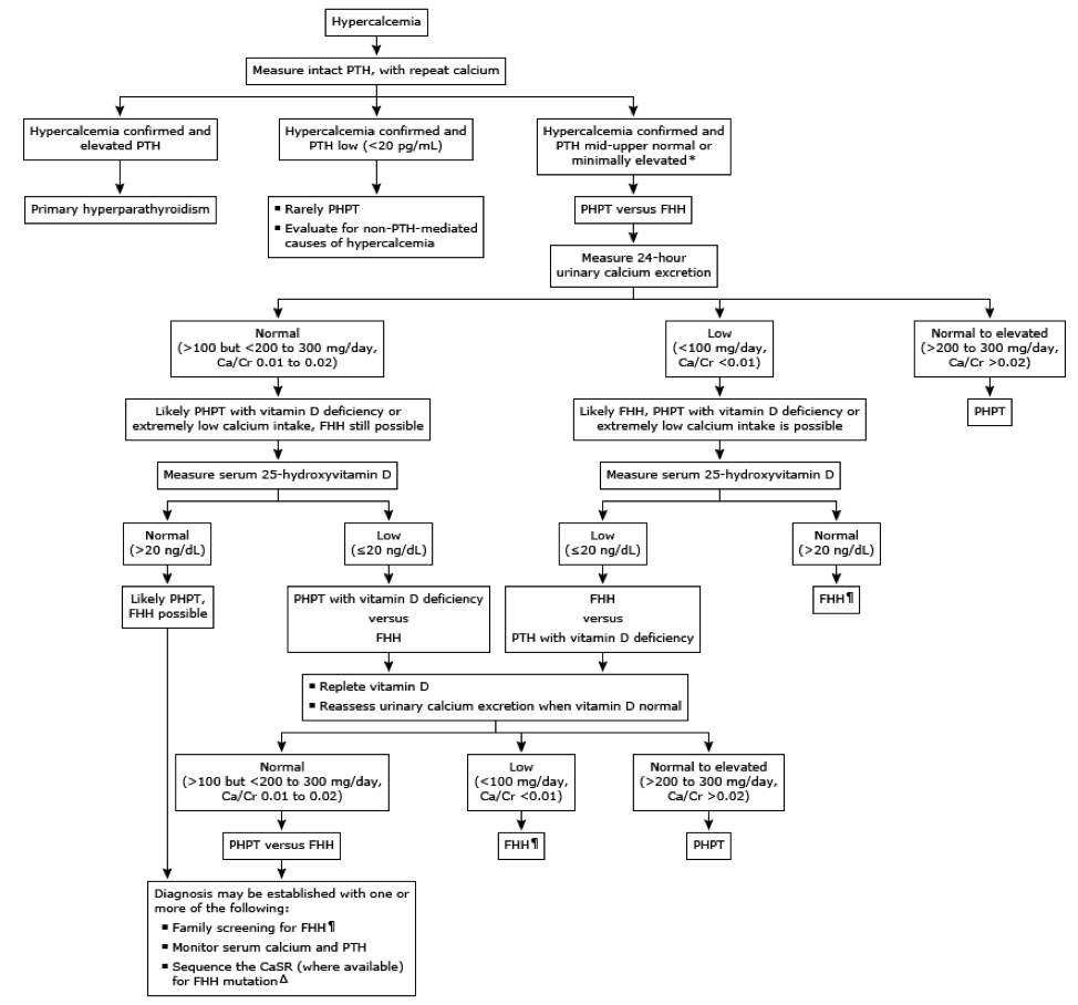
Allows for visualization of adenoma, hyperactive glands, or ectopic glands prior to surgery
Sensitivity – 70-81%, PPV – 91-95%
Procedure
Bilateral neck exploration
Medical
Bisphosphonates
Inhibits bone resorption and improves bone mass
Alendronate
Calcimimetics
Activate calcium-sensing receptors in the parathyroid gland, which decrease PTH secretion
Cinacalcet
Vitamin D
Secondary
Phosphate binders
Vitamin D supplementation
References
Potts JT, Juppner H. Parathyroid hormone: Molecular biology and regulation. In: Principles of Bone Biology, Bilezikian JP, Raisz LG, Rodan GA (Eds), Academic Press, San Diego 1996. p.325.
Diaz R, El-Hajj Fuleihan G, Brown EM. Regulation of parathyroid function. In: Handbook of Physiology, Section 7: The Endocrine System, Fray GGS (Ed), Oxford University Press, New York 1999.
Talmage RV, Mobley HT. Calcium homeostasis: reassessment of the actions of parathyroid hormone. General and Comparative Endocrinology. 2008;156(1):1-8. [pubmed]
van Abel M, Hoenderop JG, van der Kemp AW, Friedlaender MM, van Leeuwen JP, Bindels RJ. Coordinated control of renal Ca(2+) transport proteins by parathyroid hormone. Kidney International. 2005;68(4):1708-21. [pubmed]
Bartsch D, Nies C, Hasse C, Willuhn J, Rothmund M. Clinical and surgical aspects of double adenoma in patients with primary hyperparathyroidism. The British Journal of Surgery. 1995;82(7):926-9. [pubmed]
Ruda JM, Hollenbeak CS, Stack BC. A systematic review of the diagnosis and treatment of primary hyperparathyroidism from 1995 to 2003. Otolaryngology. 2005;132(3):359-72. [pubmed]
Rafferty MA, Goldstein DP, Rotstein L. Completion thyroidectomy versus total thyroidectomy: is there a difference in complication rates? An analysis of 350 patients. Journal of the American College of Surgeons. 2007;205(4):602-7. [pubmed]
Silverberg SJ, Bilezikian JP. Evaluation and management of primary hyperparathyroidism. The Journal of Clinical Endocrinology and Metabolism. 1996;81(6):2036-40. [pubmed]
Coker LH, Rorie K, Cantley L. Primary hyperparathyroidism, cognition, and health-related quality of life. Annals of Surgery. 2005;242(5):642-50. [pubmed]
Rastogi R, Beauchamp NJ, Ladenson PW. Calcification of the basal ganglia in chronic hypoparathyroidism. The Journal of Clinical Endocrinology and Metabolism. 2003;88(4):1476-7. [pubmed]
Stein R, Godel V. Hypocalcemic cataract. Journal of Pediatric Ophthalmology and Strabismus. 1980;17(3):159-61. [pubmed]
Kinirons MJ, Glasgow JF. The chronology of dentinal defects related to medical findings in hypoparathyroidism. Journal of Dentistry. 1985;13(4):346-9.[pubmed]
Goltzman D, Cole DEC. Hypoparathyroidism. In: Primer on the metabolic bone diseases and disorders of mineral metabolism, 6th ed, Favus MJ. (Ed), American Society of Bone and Mineral Research, Washington, DC 2006. p.216.
Winer KK, Ko CW, Reynolds JC. Long-term treatment of hypoparathyroidism: a randomized controlled study comparing parathyroid hormone-(1-34) versus calcitriol and calcium. The Journal of Clinical Endocrinology and Metabolism. 2003;88(9):4214-20. [pubmed]
Bilezikian JP, Brandi ML, Eastell R. Guidelines for the management of asymptomatic primary hyperparathyroidism: summary statement from the Fourth International Workshop. The Journal of Clinical Endocrinology and Metabolism. 2014;99(10):3561-9. [pubmed]
Eslamy HK, Ziessman HA. Parathyroid sctinigraphy in patients with primary hyperparathyroidism: 99mTc sestamibi SPECT and SPECT/CT. Radiographics. 2088;28:1461-1476.
What are the indications for high-intensity statin therapy for hyperlipidemia?
New joint guidelines were released in 2013 by ACC/AHA on the management of hyperlipidemia. In these guidelines, there are 3 criteria for high intensity statin therapy:
Age < 75 years with a clinical Atherosclerotic Cardiovascular Disease (ASCVD):
What are the drugs and dosing for high-intensity statin therapy?
There are only 2 medications that have been studied that are recommended for high-intensity statin therapy:
Atorvastatin 40-80mg daily
Rosuvastatin 20-40mg daily
2013 ACC/AHA Hyperlipidemia Guidelines
Knowmedge
Reference
Stone NJ, Robinson JG, Lichtenstein AH. 2013 ACC/AHA guideline on the treatment of blood cholesterol to reduce atherosclerotic cardiovascular risk in adults: a report of the American College of Cardiology/American Heart Association Task Force on Practice Guidelines. Journal of the American College of Cardiology. 63(25 Pt B):2889-934. 2014. [pubmed]