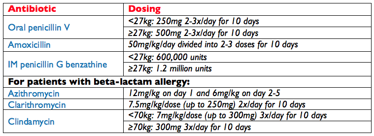
American Academy of Pediatrics. Group A Streptococcal Infections. In: Red Book: 2015 Report of the Committee on Infectious Diseases, 30th, Kimberlin DW, Brady MT, Jackson MA, Long SS (Eds), American Academy of Pediatrics, Elk Grove Village, IL 2015. p.732.
With finals week closely approaching, this weekend snuck up on me quick and I was not able to get a good case together for the blog….my bad.
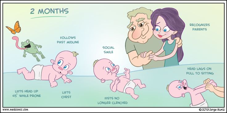
But, FEAR NOT!!!! What I thought I would do for the last post of pediatric month is review the pediatric developmental milestones by way of infographics.
If you haven’t already, you need to be following Jorge Muniz of Medcomic on Twitter and buy his book….it is awesome and he is a fellow PA. I try to incorporate as many of his images as possible when I teach.
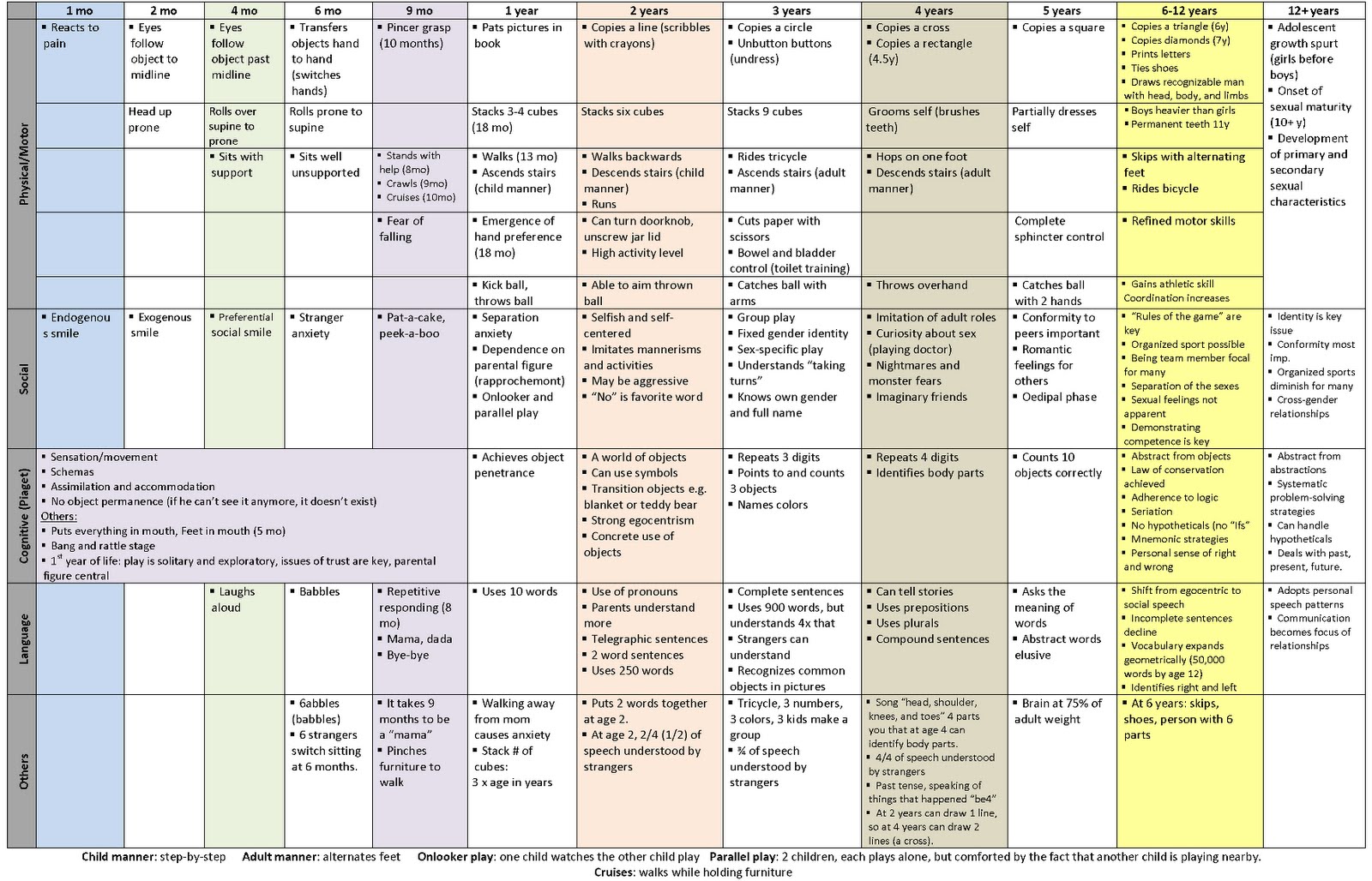
The last graphic is the THE BEST single graphic of developmental milestones (from 1 month to 12 years) I have ever found. Great resource for your pediatric rotation.
Comprise 15% of all CHD and 33% of potentially fatal CHD
Physiology
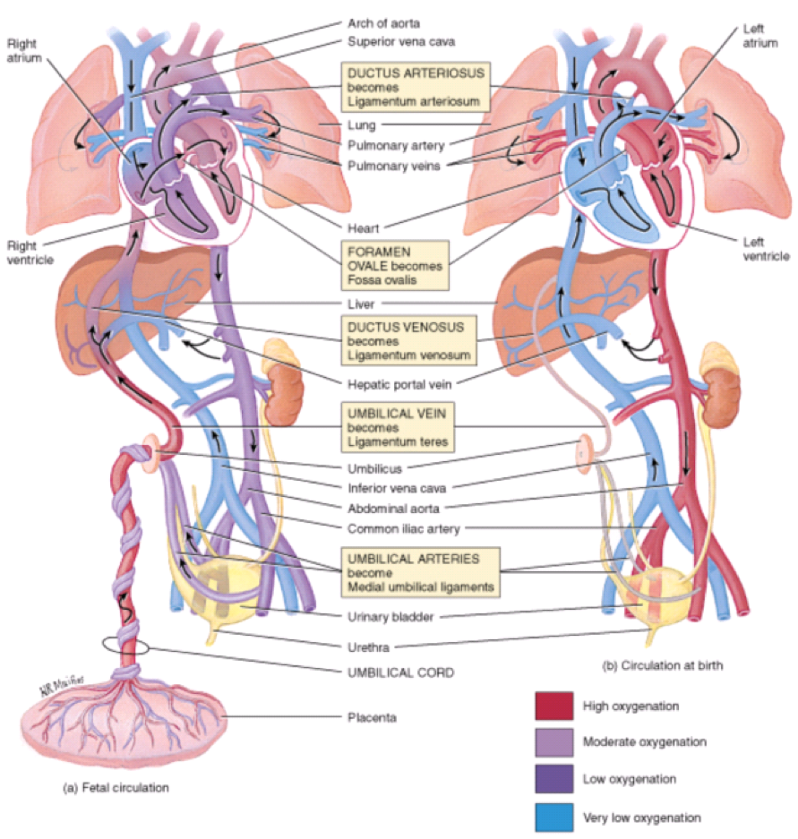
The cardiovascular system in-utero is a complicated machine that is designed to bypass the lungs and provide oxygenated blood from the placenta. There are two main structures that help maintain oxygenation when the fetus’ lungs are not used:
Ductus arteriosus
Connects the pulmonary artery to the descending aorta
Prostaglandin E1 and E2 are produced by the placenta and keep this open
Absolutely vital to remain patent in several of the cyanotic diseases to provide oxygenated blood
Foramen ovale
Communication between right and left atrium
Once the infant begins spontaneously breathing, increases in pulmonary blood flow and left atrial pressures mechanically seals the foramen ovale
Fetal circulation (a) in-utero and (b) during 1st 7 days of life
Khan Academy Tutorials
Cardiac Causes of Cyanosis
3 Main Physiologic Categories
Decreased pulmonary blood flow
Tetralogy of Fallot, tricuspid atresia
Increased pulmonary blood flow
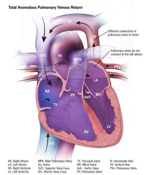
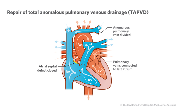
Transposition of great vessels, truncus arteriosis, total anomalous pulmonary venous connection
Severe heart failure
Hypoplastic left heart, coarctation of the aorta
Timing of Presentation
Within 48 hours of birth
Transposition of great vessels, tricuspid atresia
With 7 days of birth
Truncus arteriosus, total anomalous pulmonary venous connection, Tetralogy of Fallot
Screening
Hyperoxia Test
100% oxygen via hood for 10 minutes
Radial artery (preductal) PaO2 is measured
PaO2 > 150 mmHg suggests pulmonary disease
Pulse Oximetry Screening
Measuring the difference in SpO2 between preductal (right hand) and postductal (either foot) flow
A positive test warranting further investigation includes any of the following:
SpO2 < 90% in either extremity
SpO2 90-94% in both locations on three measurements one hour apart
SpO2 difference > 3% on three measurements one hour apart
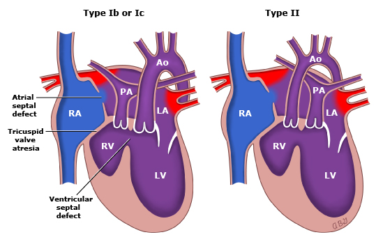
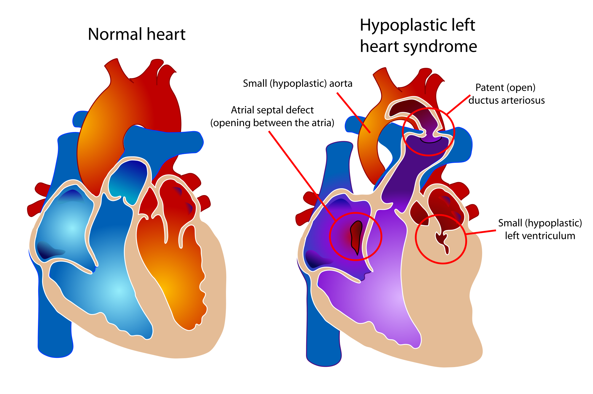
Spectrum of cardiac malformations characterized by underdevelopment of the left ventricle with atresia, stenosis, or hypoplasia of aortic and/or mitral valve, and hypoplasia of ascending aorta and arch
Survival is dependent on PDA and ASD
Signs and Symptoms
Prenatal
Can be diagnosed by fetal ultrasound between 18-24 weeks
Postnatal
“Honeymoon” period while PDA is open and ASD is unrestricted
May be discharged and present after 3-5 days
If ASD is restricted –> rapid decompensation as PDA closes
Single S2 heart sound
No murmur
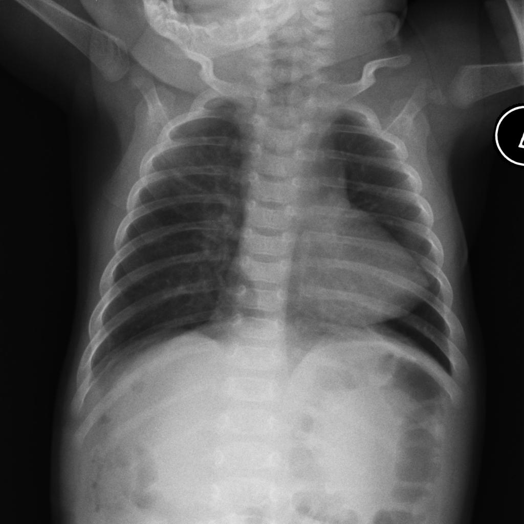
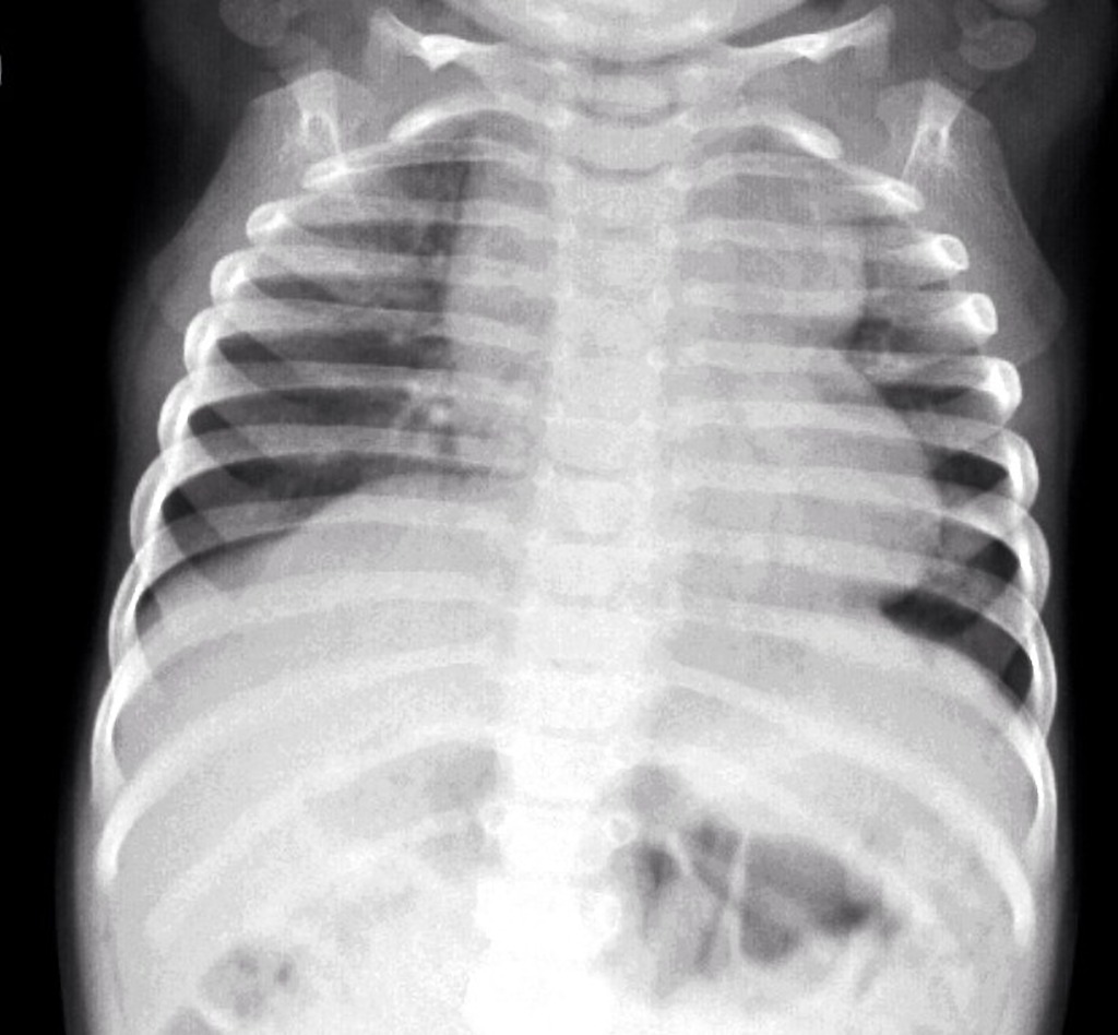
Chest radiograph may show small cardiac silhouette
Electrocardiogram shows RAD, RAE, RVH
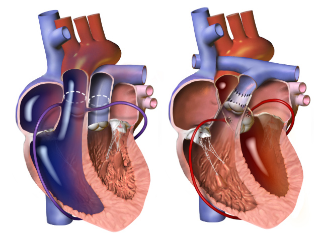
Surgical repair performed in 3 stages
1st stage performed immediately
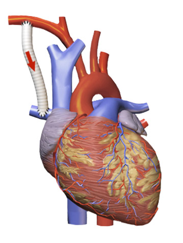
Norwood procedure (3 parts)
Creation of neoaorta
Blalock-Taussig shunt
Resection of atrial septum
Norwood Procedure
2nd stage performed at 3-6 months
Bidirectional Glenn procedure
Bidirectional Glenn Procedure
3rd stage performed at 2-3 years
Fontan procedure
Hybrid approach and heart transplant are emerging treatment options
PAINE Pearls to Remember
6 “Ts” of Congenital Cyanotic Heart Defects
Tetralogy of Fallot
Transposition of Great Vessels
Tricuspid Atresia
Truncus Arteriosus
Total Anomalous Pulmonary Venous Connection
“Tiny” (Hypoplastic) Left Heart Syndrome
Numbers of Congenital Cyanotic Heart Defects
1 trunk (truncus arteriosus)
2 great vessels (transposition)
3 “tri” (tricuspid atresia)
4 “tetra” (Tetralogy of Fallot)
5 words (Total Anomalous Pulmonary Venous Connection)
VI – the left “I” is half as big as the right “V” (hypoplastic left heart)
Cottage Physician Reference
Nothing directly related to congenital heart defects, but I did find this quote interesting. It says:
“ The general rule as to tying the cord , with the exceptions above noticed, is, that it is the safest to delay the tying of it, until it has entirely ceased to pulsate”
The OB realm is still debating delayed cord clamping…It looks like everything in medicine always comes full circle


