Ep-PAINE-nym


Fournier’s Gangrene

Other Known Aliases – none

Definitionnecrotizing fasciitis of the external genitalia and/or perineum

Clinical Significance this infection commonly affects older men and is associated with diabetes mellitus or a compromised immune system. Other risk factors include trauma or surgery to the perineal area, alcoholism, and childbirth. Pain, erythema, crepitus, and fever are common findings and treatment is aggressive surgical debridement and antibiotics to cover anaerobic and facultative pathogens.
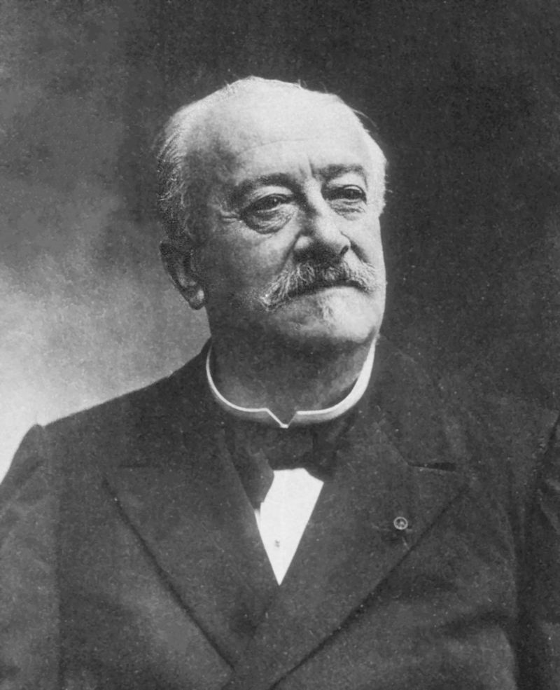
HistoryNamed after Jean Alfred Fournier (1832-1914), who was a French dermatologist and venereologist, and received his medical doctorate in 1860 while studying in Paris. He would begin his career as an understudy of Philippe Ricord at the Hôpital du Midi and would later become médecine des hôpitaux at the famed Hôtel-Dieu de Paris. It was in 1883 when he presented a case series of patients with gangrene of perineum and for which this eponym is attributed, although it was first described and published in 1764 by Baurienne. He is best known for his work with congenital syphilis (for which he has two additional eponyms) and advancing the study of venereal diseases and their connection to degenerative diseases.


References

  1. Firkin BG and Whitwirth JA.  Dictionary of Medical Eponyms. 2nd ed.  New York, NY; Parthenon Publishing Group. 1996.
  2. Bartolucci S, Forbis P.  Stedman’s Medical Eponyms.  2nd ed.  Baltimore, MD; LWW.  2005.
  3. Yee AJ, Pfiffner P. (2012).  Medical Eponyms (Version 1.4.2) [Mobile Application Software].  Retrieved http://itunes.apple.com.
  4. Whonamedit – dictionary of medical eponyms. http://www.whonamedit.com
  5. Up To Date. www.uptodate.com
  6. Fournier, J.A. Gangrène foudroyante de la verge. La Semaine Médicale. 3 1883;(56): 345–347
  7. Toodayan N. Jean Alfred Fournier (1832-1914): His contributions to dermatology. http://www.odermatol.com/issue-in-html/2015-4-32/
  8. Waugh MA. Alfred Fournier, 1832-1914. His influence on venereology. Br J Vener Dis. 1974; 50(3):232-6. [PDF]

PAINE #PANCE Pearl – Rheumatology


Question

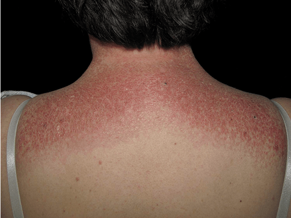
49yo woman, with a history of hypertension and GERD, presents to your clinic with a six month history of bilateral shoulder weakness that she first noticed when putting on sweaters and jackets and carrying in groceries. She denies any history of pain, repetitive trauma, or weakness in the hands. On physical examination, she has 3.5/5 strength in shoulder abduction and flexion, as well as the below rash. She reports the rash has been present for about the same time, but doesn’t really bother her.

  1. What lab is likely to be profoundly elevated and what lab is most specific to this condition?
  2. What is the most likely diagnosis?


Answer

  1. Due to the inflammatory myopathy, muscle enzymes are often extremely elevated and are helpful in initial screening. Creatine kinase (CK) is most commonly ordered, but lactate dehydrogenase (LDH), aspartate aminotransferase (AST), and alanine aminotransferase (ALT) are often elevated as well. The most common myositis-specific autoantibody is Anti-Jo 1 with others being Anti-SRP and Anti-MI-2.
  2. Given this history and dermatologic “shawl sign”, dermatomyositis is most likely. Polymyositis does not present with skin findings.

Ep-PAINE-nym


Kienböck Disease

Other Known Aliases – none

Definitionavascular necrosis of the lunate

Clinical Significance most often results from trauma with biomechanical and vascular abnormalities that lead to progressive bone death. Patients will report wrist pain with decreased range of motion and grip strength. MRI is best for early diagnosis and treatment depends on the stage of disease using the Lichtman Classification system.
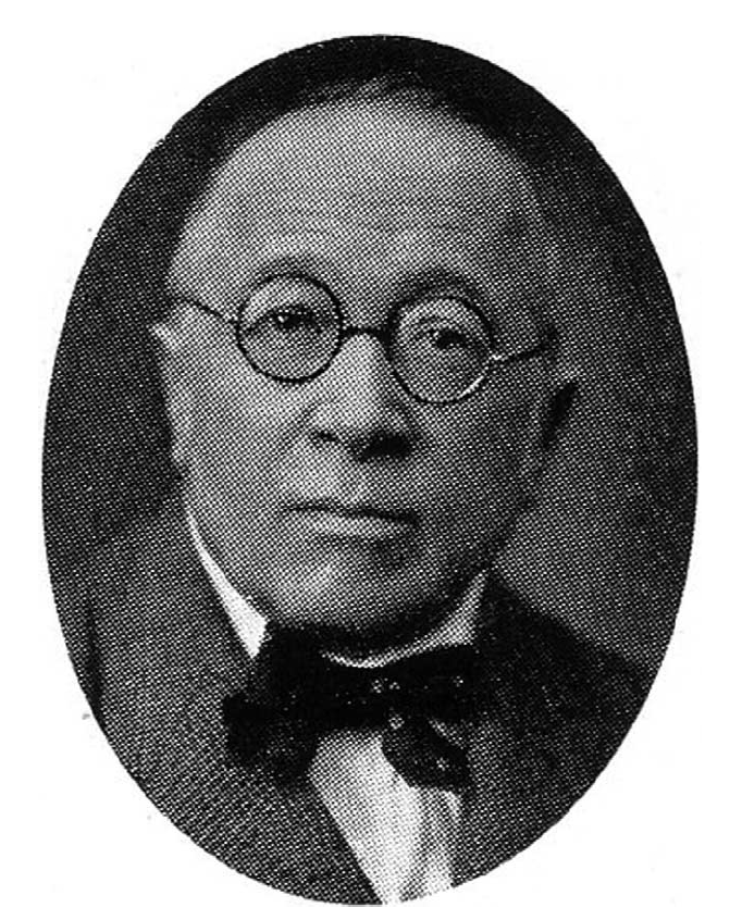
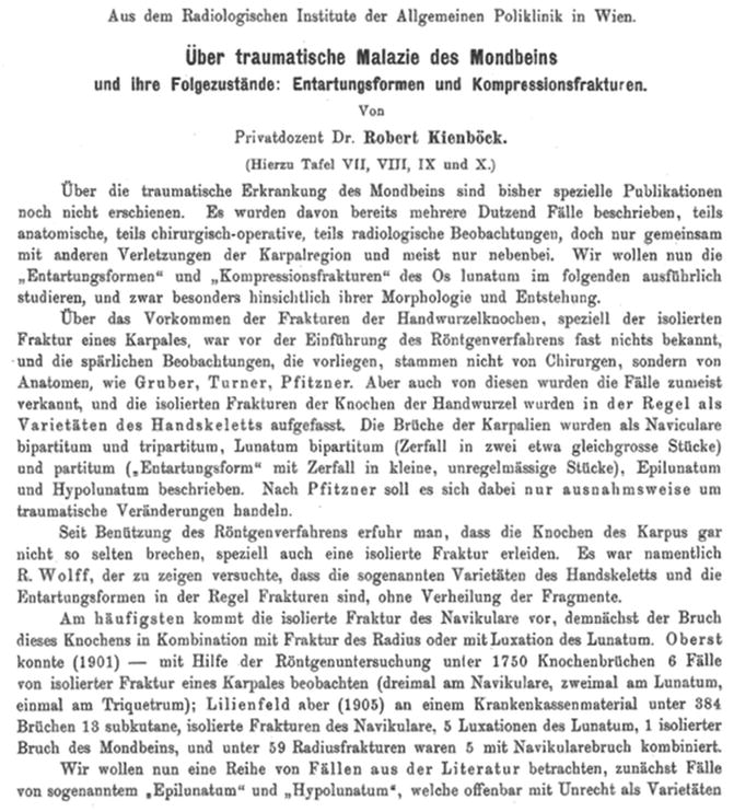
HistoryNamed after Robert Kienböck (1871-1953), who was an Austrian radiologist and received his medical doctorate from the University of Vienna in 1895. He would explore the new and blossoming field of radiology before becoming the head of the radiological department at Vienna General Hospital before becoming professor of radiology in 1926. He was a pioneer in the use of x-rays for medical diagnosis and would co-found the Vienna Radiology Society in 1923. He would publish his eponymous condition in 1910 in his treatise Über traumatische Malazie des Mondbeins und ihre Folgezustände (Traumatic malacia of the lunate and its consequences).


References

  1. Firkin BG and Whitwirth JA.  Dictionary of Medical Eponyms. 2nd ed.  New York, NY; Parthenon Publishing Group. 1996.
  2. Bartolucci S, Forbis P.  Stedman’s Medical Eponyms.  2nd ed.  Baltimore, MD; LWW.  2005.
  3. Yee AJ, Pfiffner P. (2012).  Medical Eponyms (Version 1.4.2) [Mobile Application Software].  Retrieved http://itunes.apple.com.
  4. Whonamedit – dictionary of medical eponyms. http://www.whonamedit.com
  5. Up To Date. www.uptodate.com
  6. Orthobullets. Kienbock Disease. https://www.orthobullets.com/hand/6050/kienbocks-disease
  7. Wagner JP, Chung KC. A Historical Report on Robert Kienböck and Kienböck Disease. The Journal of Hand Surgery. 2005;30(6):1117-1121. [link]
  8. Kienböck R. Über traumatische Malazie des Mondbeins und ihre Folgezustände: Entartungsformen und Kompressionfrakturen. Fortschritte auf dem Gebiete der Röntgenstrahlen. 1910–1911; 16: 77-103.

#63 – Osteoarthritis Treatment


***LISTEN TO THE PODCAST HERE***


Quick Osteoathritis Review

  • Signs and Symptoms
    • Progressive joint pain
      • Stage 1 – Intermittent, Predictable, limits only high-impact activities
      • Stage 2 – Constant, effects daily activities
      • Stage 3 – Constant with intermittent, unpredictable, intense pain with severe limitations
    • Worse in the afternoon
    • Decreased ROM
    • Joint-line tenderness
    • Swelling and effusions
  • Radiography
    • 4 Radiographic Criteria of Osteoarthritis
      • Diminished joint space
      • Bony sclerosis
      • Osteophytes
      • Subchondral cyst


General Osteoarthritis Treatment Principles

  • Education
    • Discuss modifiable risk factors
    • Prognosis
    • Treatment options and timeline
  • Goal Setting
    • Identify current issues
    • Set priorities
    • Develop realistic plan
      • Multiple short term goals to achieve long term goal
      • Directed at minimizing pain, optimizing function, and modify joint damage
  • Clinical Assessment and Follow-up
    • Should be every 3 months by provide
    • Factors to be addressed and discussed during visits:
      • Impact of pain on daily living and quality of life
      • Functional limitations
      • Recreational and/or occupational aspirations
      • Sleep disturbances
      • Fall risk assessment
      • Expectations of treatment

Updated Guidelines from ACR/AAF

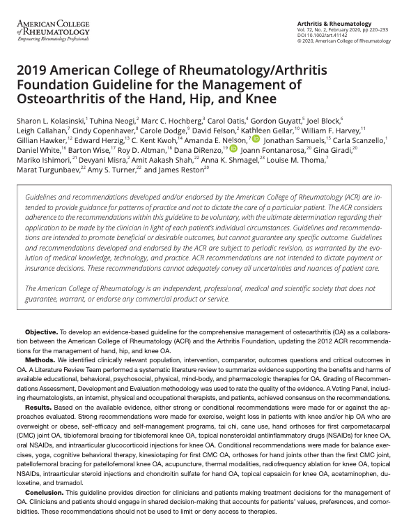
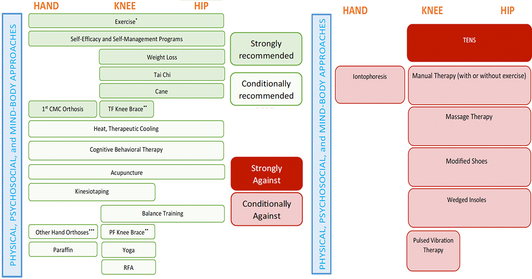
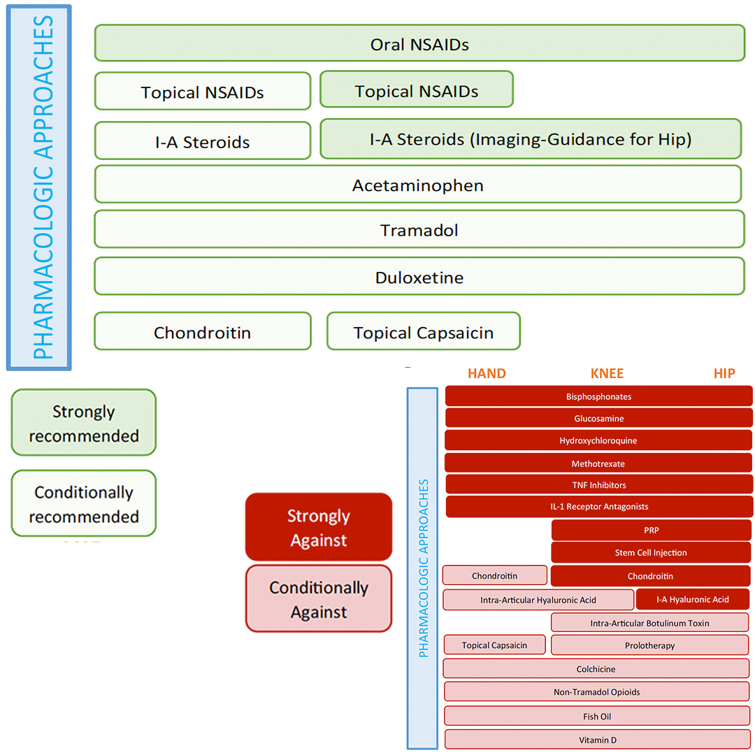
2019 updated guidelines from American College of Rheumatology and American Arthritis Foundation


Non-Pharmacologic Treatment

  • Should be first line either alone or with pharmacologic therapy
  • Physical therapy is the mainstay of non-pharmacologic treatment
    • Usually 6 weeks
  • Weight loss
    • Loss of 10% of body weight equals 50% reduction in pain scores
    • Adipokines (leptin and adiponectin) released by adipose tissue are known inflammatory factors
    • Consultation with dietician can be helpful
  • Exercise
    • Exercising have comparable effects on pain and function compared to NSAIDs
    • Low-impact is best, but tailor to patient’s function and limitations
    • Activities to help with core strength and balance can have significant reductions in falls
  • Braces and Splints
    • When possible, these aids can have significant benefit in pain reduction during activities

Pharmacologic Treatments

  • Topical NSAIDs should be considered prior to oral NSAIDs
    • Knee > hand efficacy
    • Diclofenac 1% gel – 4g (large joints) or 2g (small joints) applied 3-4x/day
      • Now available OTC
  • Oral NSAIDs > acetaminophen
    • COX-2 selective NSAID
      • Celecoxib 100-200mg daily or BID
      • Diclofenac 75mg BID
      • Meloxicam 10-15mg daily
  • Duloxetine can be helpful
    • Desensitizes central nociceptive pain processing
    • 60-120mg daily
  • Intrarticular glucocorticoid injections have limited role
    • Can be used for short-term relief
      • Most helpful with the hip
      • Long-term use can damage cartilage
    • Triamcinolone 40mg
  • Hyaluronic acid is controversial with limited data
  • Avoid recommended glucosamine, chondroitin, vitamin D, and fish oil due to lack of clear data showing benefit

Surgical Indications and Management


Cottage Physician (1893)


References

  1. Hawker GA, Stewart L, French MR, et al. Understanding the pain experience in hip and knee osteoarthritis–an OARSI/OMERACT initiative. Osteoarthritis Cartilage. 2008; 16(4):415-22. [pubmed]
  2. French SD, Bennell KL, Nichols PJ, Hodges PW, Dobson FL, Hinman RS.  What do people with knee or hip osteoarthritis need to know? An international consensus of essential statements for osteoarthritis.  Arthritis Care Res (Hoboken). 2015;57(6):809. [pubmed]
  3. Bodenheimer T, Lorig K, Holman H, Grumbach K. Patient self-management of chronic disease in primary care. JAMA. 2002; 288(19):2469-75. [pubmed]
  4. Messier SP, Mihalko SL, Legault C, et al. Effects of intensive diet and exercise on knee joint loads, inflammation, and clinical outcomes among overweight and obese adults with knee osteoarthritis: the IDEA randomized clinical trial. JAMA. 2013; 310(12):1263-73. [PDF]
  5. Zhang W, Nuki G, Moskowitz RW, et al. OARSI recommendations for the management of hip and knee osteoarthritis: part III: Changes in evidence following systematic cumulative update of research published through January 2009. Osteoarthritis Cartilage. 2010; 18(4):476-99. [pubmed]
  6. 2019 American College of Rheumatology/Arthritis Foundation Guideline for Management of Osteoarthritis of the Hand, Hip, and Knee.  Arthritis Care Res (Hoboken). 2020;72(2):149-162. [pubmed]

PAINE #PANCE Pearl – Rheumatology


Question

43yo woman, with a history of hypertension and GERD, presents to your clinic with a six month history of bilateral shoulder weakness that she first noticed when putting on sweaters and jackets and carrying in groceries. She denies any history of pain, repetitive trauma, or weakness in the hands. On physical examination, she has 3.5/5 strength in shoulder abduction and flexion, as well as the below rash. She reports the rash has been present for about the same time, but doesn’t really bother her.

  1. What lab is likely to be profoundly elevated and what lab is most specific to this condition?
  2. What is the most likely diagnosis?

Ep-PAINE-nym


Monteggia Fracture

Other Known Aliasesnone

Definitionproximal 1/3 ulnar fracture with radial head dislocation

Clinical Significance This type of injury pattern is most commonly seen with FOOSH injuries and is more common in children than adults with a peak incidence of 4-10 years of age. There are four different classifications depending on the injury pattern. There is also high incidence of neurovascular compromise and a good bedside exam is paramount prior to surgical repair.

HistoryNamed after Giovanni Battista Monteggia (1762-1815), who was an Italian surgeon and received his medical doctorate from the University of Pavia in 1789 at the age of seventeen. He would begin his career as a surgery apprentice at the Great Hospital in Milano in 1790 culminating in professor of anatomy and surgery in 1795. His knowledge of anatomy and skill as a surgeon impressed his a very famous colleague at the University of Pavia, one Antonio Scarpa. He published his eponymous injury in 1814 in his textbook entitled “Institziono Chirurgiche”. Of note, the first radiograph was not taken until 1895.


References

  1. Firkin BG and Whitwirth JA.  Dictionary of Medical Eponyms. 2nd ed.  New York, NY; Parthenon Publishing Group. 1996.
  2. Bartolucci S, Forbis P.  Stedman’s Medical Eponyms.  2nd ed.  Baltimore, MD; LWW.  2005.
  3. Yee AJ, Pfiffner P. (2012).  Medical Eponyms (Version 1.4.2) [Mobile Application Software].  Retrieved http://itunes.apple.com.
  4. Whonamedit – dictionary of medical eponyms. http://www.whonamedit.com
  5. Up To Date. www.uptodate.com
  6. Orthobullets. Monteggia Fractures. https://www.orthobullets.com/trauma/1024/monteggia-fractures
  7. Monteggia GB. Instituzioni Chirurgiches. Vol. 5. Maspero; Milan, Italy: 1814.

PAINE #PANCE Pearl – Orthopaedics


Question

57yo woman comes into clinic complaining of a six-month history of right shoulder pain. She states that the pain is worse with overhead activities of the shoulder and when she is lifting and pulling with that arm. She has been more playing more tennis with her husband during this time as well, but has never had any other issues with this shoulder or arm before. She also reports painful arc of shoulder rotation with clicking that she feels on the anterior part of her shoulder. On physical examination, she has a negative drop arm and empty can test with no significant pain on resisted external rotation.

  1. What are two (2) maneuvers that you should perform next?
  2. If these are positive, what would the most likely diagnosis be?


Answer

Given the history and negative findings on rotator cuff maneuvers, the next two maneuvers shoulder be the Yergason and Speed tests. If the patient has pain with either of these then it suggests biceps tendonitis as the cause of their shoulder pain. Anatomically, the origin of the long head of the biceps is near the insertion of the supraspinatus tendon. This is why you always assess biceps tendonopathy in patient presenting with shoulder pain.

Ep-PAINE-nym


De Quervain Tenosynovitis

Other Known AliasesBlackBerry thumb, mother’s wrist, washerwoman’s sprain

Definitioninflammation of the abductor pollicis longus and extensor pollicis brevis tendons in the first extensor compartment at the styloid process of the radius.

Clinical Significance The pathogenesis is not well understood, but is most accepted to be caused by repetitive activities that maintain the thumb in extension and abduction. Treatment is graded from thumb spica splinting, NSAIDs, corticosteroid injections, up to surgical release of the first extensor compartment.

HistoryNamed after Fritz de Quervain (1868-1940), who was a Swiss surgeon and received his medical doctorate from the University of Bern in 1892. He would start his career training under Hugo Kronecker, Theodor Langhans, and Theodor Kocher, before becoming director of the surgical department at the La Chaux-de-fonds in Neuchâtel in 1897, and culminating in professor of surgery and director of the Inselspital at the University of Bern in 1918. He was a strong proponent of the generalist approach to patient care rather than the specialization of physicians and surgeons. He would describe his eponymous findings in his classic textbook series Spezielle chirurgische Diagnostik (Special Surgical Diagnosis) in 1907.


References

  1. Firkin BG and Whitwirth JA.  Dictionary of Medical Eponyms. 2nd ed.  New York, NY; Parthenon Publishing Group. 1996.
  2. Bartolucci S, Forbis P.  Stedman’s Medical Eponyms.  2nd ed.  Baltimore, MD; LWW.  2005.
  3. Yee AJ, Pfiffner P. (2012).  Medical Eponyms (Version 1.4.2) [Mobile Application Software].  Retrieved http://itunes.apple.com.
  4. Whonamedit – dictionary of medical eponyms. http://www.whonamedit.com
  5. Up To Date. www.uptodate.com
  6. Spezielle chirurgische Diagnostik. Leipzig, 1907; 9th edition, 1931.

PAINE #PANCE Pearl – Orthopaedics


Question

57yo woman comes into clinic complaining of a six-month history of right shoulder pain. She states that the pain is worse with overhead activities of the shoulder and when she is lifting and pulling with that arm. She has been more playing more tennis with her husband during this time as well, but has never had any other issues with this shoulder or arm before. She also reports painful arc of shoulder rotation with clicking that she feels on the anterior part of her shoulder. On physical examination, she has a negative drop arm and empty can test with no significant pain on resisted external rotation.

  1. What are two (2) maneuvers that you should perform next?
  2. If these are positive, what would the most likely diagnosis be?