Ep-PAINE-nym


Homan’s Sign

Other Known Aliasesdorsiflexion sign

Definitionpain in the posterior leg (classically behind the knee) with forced dorsiflexion of the foot

Clinical Significancethis examination finding was used in patients with a suspected DVT and before D-Dimers and clinical ultrasound were readily available. It is clinically useless as it has been studied extensively and found to have a sensitivity of 10-54% and specificity of 29-89%, thus not ruling in or out the condition consistently.
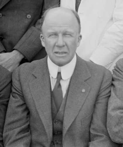
HistoryNamed after John Homans (1877-1954), who was an American surgeon and received his medical doctorate from Harvard Medical School. He worked with Harvey Cushing and Samuel Crowe early in career exploring the connection between the piuitary gland and the reproductive system. He first described his eponymous finding in 1944 in a NEJM article entitled “Diseases of the veins” and later published the first case report of a DVT occuring after prolonged sitting on a flight between Boston and Caracas in 1954. He was a founding member of the the Society for Vascular Surgery and the namesake of the John Homans Chair of Surgery position at Harvard Medical School and John Homans Fellowship in Vascular Surgery at the Brigham and Women’s Hospital.


References

  1. Firkin BG and Whitwirth JA.  Dictionary of Medical Eponyms. 2nd ed.  New York, NY; Parthenon Publishing Group. 1996.
  2. Bartolucci S, Forbis P.  Stedman’s Medical Eponyms.  2nd ed.  Baltimore, MD; LWW.  2005.
  3. Yee AJ, Pfiffner P. (2012).  Medical Eponyms (Version 1.4.2) [Mobile Application Software].  Retrieved http://itunes.apple.com.
  4. Whonamedit – dictionary of medical eponyms. http://www.whonamedit.com
  5. Up To Date. www.uptodate.com
  6. McGee, Steven (2012). Evidence-Based Physical Diagnosis. Philadelphia, USA: Saunders. pp. 472–473. ISBN978-1-4377-2207-9
  7. Homans J. Diseases of the veins. N Engl J Med 1944: 231; 51-60
  8. Homans J. Thrombosis of the deep leg veins due to prolonged sitting. The New England journal of medicine. 1954; 250(4):148-9. [pubmed]
  9. Barker WF. John Homans, MD, 1877-1954. Arch Surg. 1999;134(9):1019–1020. doi:10.1001/archsurg.134.9.1019

PAINE #PANCE Pearl – Hematology


Question

57yo Caucasian male presents to his primary provider with a one-year history of joint pain, weakness, and fatigue. He has a past medical history significant for hypertension and hyperlipidemia, for which he is being treated and is controlled on medications. His wife reports his skin has become a little darker over the last year as well. The rest of his physical examination does not reveal any abnormalities. Routine chemistries show a glucose of 214 mg/dL, AST of 472 mg/dL, and ALT of 513 mg/dL. What two (2) laboratory studies should be ordered next?

Ep-PAINE-nym


Schilling Test

Other Known Aliases – none

Definitionlaboratory test for pernicious anemia (specifically intrinisic factor deficiency) that led to vitamin B12 (cobalamin) deficiency. It involved ingesting a oral dose of radiolabeled vitamin B12 (to test oral absorption), an IM injection of vitamin B12 (to saturate liver stores), and a 24hr urine collection to see how much was absorbed and excreted. If intestinal absorption was intact (intrinsic factor present), then > 10% of the radiolabeled vitamin B12 would be in the urine.

Clinical SignificanceThis was the first and only test at the time to be able to diagnose pernicious anemia, but is now largely a test of historical interest only as better diagnostic studies have been developed.

HistoryNamed after Robert F. Schilling (1919-2014) an American physician and researcher who received his medical doctorate from the University of Wisconisn-Madison in 1943. Immediately after graduation, he joined the Pacific Front as a physician in the 3d Marine Division. After the war, he completed postgraduate training at Harvard before returning to Wisconsin to practice hematology. He studied extensively on the metabolism of vitamin B12 and the urinary excretion of radiolabeled vitamin B12 in pernicious anemia and in 1953, published a paper entitled “The effect of gastric juice on the urinary excretion of radioactivity after the oral administration of radioactive vitamin B12”, which would go on to be called the “Schilling Test”.


References

  1. Firkin BG and Whitwirth JA.  Dictionary of Medical Eponyms. 2nd ed.  New York, NY; Parthenon Publishing Group. 1996.
  2. Bartolucci S, Forbis P.  Stedman’s Medical Eponyms.  2nd ed.  Baltimore, MD; LWW.  2005.
  3. Yee AJ, Pfiffner P. (2012).  Medical Eponyms (Version 1.4.2) [Mobile Application Software].  Retrieved http://itunes.apple.com.
  4. Whonamedit – dictionary of medical eponyms. http://www.whonamedit.com
  5. Up To Date. www.uptodate.com
  6. Schilling RF. Intrinsic factor studies. 2. The effect of gastric juice on the urinary excretion of radioactivity after the oral administration of radioactive vitamin B12. J Clin Lab Med. 1953;42;860-866
  7. Schilling Test. Stat Pearls. 2018. https://www.ncbi.nlm.nih.gov/books/NBK507784/

PAINE #PANCE Pearl – Oncology

Question


27yo male, with stage II Hodgkin’s lymphoma, presents to his oncologists’s office for 6-week follow-up s/p 4 cycles of ABVD chemotherapy.  He reports malaise and fatigue, but is otherwise in good spirits.  Vitals show:

  • BP – 122/72 mmHg
  • HR – 96 bpm
  • RR – 15 bpm
  • O2% – 100% on room air
  • Temperature – 38.5oC (101.4oF)


Labs in the office are below:


Answer


This patient has a neutropenic fever.  This is an oncologic emergency because their immune systems has been completely destroyed by the chemotherapy and they are unable to mount an appropriate response to the infectious insult. In order to determine if a patient is neutropenic, you must calculate an Absolute Neutrophil Count (ANC).

MD Calc Link

Neutropenia =  ANC < 1500


Ep-PAINE-nym


Hodgkin’s Lymphoma


Other Known Aliasesnone


Definition – Type of lymphoma predominantly from lymphocytes that arise from germinal center or post-germinal center of B cells


Clinical SignificanceAccounts for 10% of all lymphomas and 0.6% of all cancers.  It is also associated with a bimodal age distribution of young adults (20s) and older adults (60s), with a slight male predominance.  Epstein-Barr virus is the most common causative agent and it carries a favorable prognosis.


History – Named after Thomas Hodgkin (1798-1866), who was British physician and considered one of the most prominent pathologists of his time.  He was also a perpetual student constantly learning new techniques including being an early adopter of the stethoscope anda fervent advocate for preventative medicine.  He first described his findings on his eponymous disease in 1832, but it wasn’t until 33 years later when another British physician, Samuel Wilks, “re-discovered” the disease and Hodgkin’s work did it gain any traction and recognition.


Thomas Hodgkin photo.jpg


References

  1. Firkin BG and Whitwirth JA.  Dictionary of Medical Eponyms. 2nd ed.  New York, NY; Parthenon Publishing Group. 1996.
  2. Bartolucci S, Forbis P.  Stedman’s Medical Eponyms.  2nd ed.  Baltimore, MD; LWW.  2005.
  3. Yee AJ, Pfiffner P. (2012).  Medical Eponyms (Version 1.4.2) [Mobile Application Software].  Retrieved http://itunes.apple.com.
  4. Whonamedit – dictionary of medical eponyms. http://www.whonamedit.com
  5. Hodgkin Lymphome.  British Medical Journal – Best Practice. http://bestpractice.bmj.com/topics/en-us/311
  6. Hodgkin T.  On Some Morbid Appearances of The Absorbent Glands and Spleen.  Med Chir Trans.  1832;17:68-114.

PAINE #PANCE Pearl – Oncology


27yo male, with stage II Hodgkin’s lymphoma, presents to his oncologists’s office for 6-week follow-up s/p 4 cycles of ABVD chemotherapy.  He reports malaise and fatigue, but is otherwise in good spirits.  Vitals show:

  • BP – 122/72 mmHg
  • HR – 96 bpm
  • RR – 15 bpm
  • O2% – 100% on room air
  • Temperature – 38.5oC (101.4oF)


Labs in the office are below:


Ep-PAINE-nym


Auer Rods


Other Known Aliasesnone


Definition – Auer rods are azurophilic granules found in the cytoplasm of leukemic blast cells and are composed of fused lyosomes.


Image result for auer rods


Clinical SignificanceThese are found in high grade myelodysplastic and myeloproliferative syndromes and are pathognomonic for acute myelogenous leukemia (AML).


Image result for auer rods


History – Named after John Auer (1875-1948), an American physiologist and pharmacologist, who held appointments at Johns Hopkins Hospital, Rockefeller Institute for Medical Research, and the St. Louis School of Medicine. He first described these structures in a 21yo male who was suffering from a sore throat and nosebleed and admitted to Sir William Osler’s service for work-up.  He published this finding in 1906, but were first described by a colleague of his at Johns Hopkins Hospital, Thomas McCrae.  Interestingly, they both erroneously thought that the cells containing these structures were lymphoblasts, not myeloblasts.


Image result for john auer rods

scanned image of page 404


References

  1. Firkin BG and Whitwirth JA.  Dictionary of Medical Eponyms. 2nd ed.  New York, NY; Parthenon Publishing Group. 1996.
  2. Bartolucci S, Forbis P.  Stedman’s Medical Eponyms.  2nd ed.  Baltimore, MD; LWW.  2005.
  3. Yee AJ, Pfiffner P. (2012).  Medical Eponyms (Version 1.4.2) [Mobile Application Software].  Retrieved http://itunes.apple.com.
  4. Whonamedit – dictionary of medical eponyms. http://www.whonamedit.com
  5. Gordon SW, Krystal GW. Auer Rods. NEJM. 2017; 376(21):2065. [pubmed]
  6. Auer J. Some hitherto Undescribed Structures Found in the Large Lymphocytes of a Case of Acute Leukemia. AJMS. 1906;131(6):1002-1014 [article]
  7. McCrae T. Acute Lymphatic Leukemia, with Report of Five Cases. BMJ. 1905; 1(2304):404-8. [pubmed]
  8. John Auer. http://www.iqb.es/historiamedicina/personas/auer.htm

#29 – Review of Transfusion Medicine with Michelle Brown


***LISTEN TO THE PODCAST HERE***


Guest

Michelle Brown, PhD, MS, MLS(ASCP)SBB

Assistant Professor and Director, Healthcare Simulation Graduate Program

University of Alabama at Birmingham


Transfusion Statistics

According to the American Red Cross:

  • Someone in the US needs blood every 2 seconds
  • 36,000 units PRBC, 10,000 units of FFP, and 7,000 units of platelets are transfused every day in the US
  • Over 21 million blood components are transfused each year in the US, but only 13.6 million are collected

Process of Separation

When a person donates blood, 1 pint of whole blood is removed and then needs to be separated into its components for longer shelf life and targeted treatments.  There are 2 processes on how this occurs:

  • Platelet-Rich Process (PRP)
    • Easier, cheaper, but plasma/platelet yield is lower
    • BC Method
      • Complicated, but higher yield of plasma/platelet
  • Once the separation occurs, then the components can be treated with additives to help with viability and longevity. In the case of FFP and cryo, it is frozen to maintain potency of coagulation factors.


Packed Red Blood Cells

  • Facts
    • One unit of PRBC = 300cc
    • Hematocrit = 55-80%
    • One of PRBC should raise hemoglobin by 1g/dL and hematocrit by 3%
  • Storage Considerations
    • Can be stored for up to 42 days
    • Treated with citrate to prevent clotting
  • Indications for Transfusion
    • Recommendations from 2016 AABB Guidelines
      • ***To pair with patient symptoms***
      • Hemodynamically-stable, restrictive threshold = Hgb < 7g/dL
      • Orthopaedic surgery, cardiac surgery, or patients with CV = Hgb < 8g/dL
  • Complications
    • Hypocalcemia
    • Transfusion reactions
    • Alloimmunization

Fresh Frozen Plasma

  • Facts
    • 1 unit of FFP = 250cc
    • Contains all coagulation factors
    • Has an INR ~ 1.6
  • Storage Considerations
    • Frozen to -18-30oC within 8 hours of collection
      • Takes 10-30 minutes to thaw
    • Properly stored for up to 1 year
    • Must be used within 5 days after thawing
  • Indications for Transfusion
    • Vitamin K factor deficiency
      • Supratherapeutic vitamin K antagonist therapy
      • Liver disease
      • Massive transfusion protocol
    • DIC
    • TTP
  • Not indicated for hypovolemia and low BP
  • Complications
    • Male only plasma à decreases in TRALI

Cyoprecipitate

  • Facts
    • Precipitant that forms after FFP is frozen and thawed at 4oC
    • 1 unit of cryoprecipitate = 10-20cc
    • Contains fibrinogen, factor VIII, XIII, and vWF
  • Storage
    • Re-frozen at -18oC
      • Takes 10-30 minutes to thaw
    • Stored for up to 1 year
    • Only good for 4 hours once thawed and pooled, so order only when you need to transfuse
  • Indications for Transfusion
    • Low fibrinogen due to:
      • DIC
      • Liver disease
      • Uremia
      • Inherited disorders of fibrinogen
  • Complications
    • Same as other blood products

Platelets

  • Facts
    • 2 collection types
      • Pooled
        • Centrifuged down from whole blood
        • Combined with other donors
      • Apheresis (single donor)
        • Platelets collected from donor and RBC and plasma returned
        • Allows matching
      • Volume ~300cc
      • Six-pack of pooled or one unit of apheresed platelet should raise platelet count by 30-60,000/uL
  • Storage
    • Stored at room temperature because cold induces clustering of vWF receptor
    • Only a 5-day shelf life
  • Indications for Transfusion
    • Actively bleeding patients with thrombocytopenia
    • Prevention of spontaneous bleeding
      • Varies depending on clinical condition
      • < 10,000/uL
      • No longer recommended to transfuse prophylactically when patient is on bypass – only is patient exhibits bleeding w/ thrombocytopenia or plt dysfunction
    • Preparation for invasive procedures:
      • Neurosurgery or ocular surgery – 100,000/uL
      • Most major surgery – 50,000/uL
      • Central line placement – 20,000/uL
      • Lumbar puncture – 50,000/uL
      • Epidural anesthesia – 80,000/uL
  • Complications
    • Increased infection risk


Component Modification Techniques

Leukoreduction

  • Indicated for:
    • Chronically transfused patients
    • Solid organ or bone marrow transplant patients
    • Previous febrile non-hemolytic reaction

Irradiated

  • Irradiation inactivates lymphocytes
  • Decreased risk of Graft-versus-host disease in immunocompromised patients
  • Reduces shelf life to 28 days

Washed

  • Rinsing serum proteins away prior to transfusions
  • Indicated for severe or recurrent allergic reactions and IgA deficiency
  • Indicated if a patient (especially neonate) is hyperkalemic

CMV-Negative

  • Indicated for immunocompromised patients

Transfusion Related Complications


References

  1. American Red Cross. Blood Facts and Statistics.  http://www.redcrossblood.org/learn-about-blood/blood-facts-and-statistics.  Accessed 03/23/2017.
  2. Brown MR, Jennings PR. Avoiding overtransfusion: an update on risks and latest indications. JAAPA. 2012;25(8):42-5. [pubmed]
  3. Basu D, Kulkarni R. Overview of blood components and their preparation.  Indian J Anaesth.  2014;58(5):529-537. [pubmed]
  4. Carson JL, Guyatt G, Heddle NM. Clinical Practice Guidelines From the AABB: Red Blood Cell Transfusion Thresholds and Storage. JAMA. 2016;316(19):2025-2035. [pubmed]
  5. Life In The Fastlane. Fresh Frozen Plasma.  https://lifeinthefastlane.com/ccc/fresh-frozen-plasma-ffp/.  Accessed 03/23/2017.
  6. Roback JD, Caldwell S, Carson J. Evidence-based practice guidelines for plasma transfusion. Transfusion. 2010;50(6):1227-39. [pubmed]
  7. Kaufman RM, Djulbegovic B, Gernsheimer T. Platelet transfusion: a clinical practice guideline from the AABB. Annals of internal medicine. 2015;162(3):205-13. [pubmed]
  8. Sharma S, Sharma P, Tyler LN. Transfusion of blood and blood products: indications and complications. American family physician. 2011;83(6):719-24. [pubmed]
  9. Takpradit K. Rational use of Blood Components.    https://www.slideshare.net/tarlabgab/rational-use-of-blood-component/16.  Accessed 03/23/2017.