#42 – Postoperative Fever


***LISTEN TO THE PODCAST HERE***


My JAAPA article from October 2016 (JAAPA. 2016;29(10):23-28)


Introduction

Postoperative fever an elevation of body temperature to ≥ 38.3oC (100.4oF) following major surgical procedures.  The vast majority of postoperative fevers occur within the first 5 days of surgery, which is why the timing of the fever is so important in differentiating the potential causes.


Physiologic Response to Surgery

Surgery cause a massive inflammatory cascade due to the physiologic and cellular damage injury.  This is a completely normal homeostatic response, but the release of IL-1, IL-6, and TNF-alpha (to name a few) are all pyrogenic cytokines that can contribute to a febrile response.

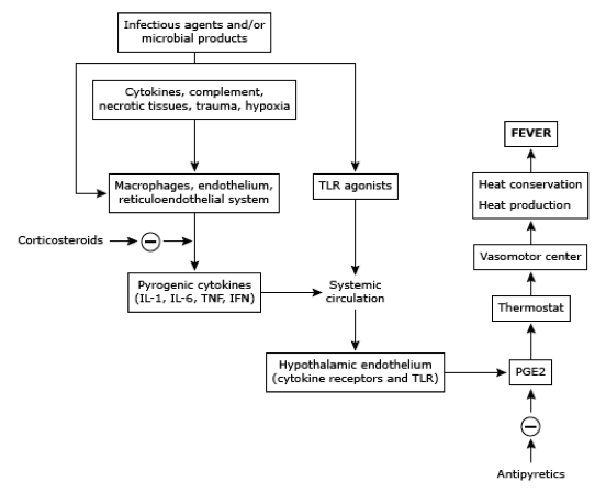
These cytokines act on the preoptic area of the hypothalamus to increase prostaglandin E2 secretion and raises the thermostatic endpoint of the body.  This cascade usually peaks at 48 hours and care should be given to prevent costly and unnecessary infectious work-ups.


The Seven W’s of Postop Fever


Wind

Atelectasis usually occurs within the first 48 hours due to splinting and incomplete lung expansion secondary to pain following surgery.  It has long been considered the leading cause of postop fever, but has been debunked in the literature for decades now with increased knowledge of the cellular inflammatory cascade.  Decreased breath sounds , SOB, crackles, tachypnea can be indicators for atelectasis and CXR may show dependent, L>R infiltrates.  Treatment should be incentive spirometry and early mobilization to promote expansion.


Water

Urinary tract infections (UTI) are the most common postoperative nosocomial infection secondary to the placement of a urinary catheter for surgery.  Risk factors for postoperative UTI are length of catheterization need, female gender, older age, history of diabetes, and previous history of UTIs.  Signs and symptoms can reveal suprapubic pain, CVA tenderness, flank pain, and malodorous, cloudy urine.  Clean catch sample is ideal, but a clean aspirate from an indwelling catheter may be used.  Urinalysis can provide a quick idea if it is infectious are not, specifically looking for positive nitrites and/or leukocyte esterase, or sending for a urine culture and finding > 105 cfu of the offending organism.  Treatment should be removing the catheter (if possible) and tailoring antibiotic therapy to the organism in question.


Wound

Surgical site infections (SSI) generally begin to manifest 3-10 after surgery and risk factors for developing are ASA score of 3-5, contaminated or dirty procedure, or an extended length of surgery.  Signs and symptoms of an SSI are increased pain, redness, and swelling at or near the incision.  These can be superficial skin, deep skin, or deep space and ultrasound can help differentiate between cellulitis and focal infection.  Treatment is either antibiotics (for cellulitis) or drainage (for abscess).


Walking

Deep venous thrombosis (DVT) can be common in surgical patient (up to 20%) secondary to immobilization and resistance to move due to pain.  To make matters worse, many can be asymptomatic with the exception of the fever.  The incidence is highest 3-5 days after surgery and you should have a low threshold to order a bilateral lower extremity ultrasound.  Treatment for a DVT following surgery can be tricky due to bleeding risk.


Wonder Drugs

Medications are the most common, noninfectious cause of fever in patients following surgery.  Timing is variable from immediately after administration to hours, or even days, later.  Most cases are due to antibiotics or heparin and result in only a mild, transient febrile response.  There are 3 life-threatening cause of medication induced fevers that you at least need to think of and rule-out:

  • Serotonin syndrome
  • Malignant hyperthermia
  • Neuroleptic malignant syndrome


Withdrawal

Up to 50% of adults over the age of 18 admit to regular alcohol consumption and up to 50% of these patients report withdrawal symptoms when abstinent.  Signs and symptoms of acute withdrawal can begin to manifest 6-72 hours after the last ingestion and range in severity from mild, vague symptoms to florid delirium tremens.  Treatment is aggressive benzodiazepine regimens to abate the symptoms, with a gradual taper while in the hospital.


“Wonky” Glands

Though rare, two endocrinologic conditions can cause fever in the postoperative period.  Adrenal insufficiency and thyrotoxicosis can both occur due to disruption of the hypothalamic-pituitary-adrenal/thyroid axis by the inflammatory mediated response of surgery.  Coupled with a patient being NPO prior to surgery, a patient may not be able to take their medications to keep these conditions at bay.  Careful preoperative history will identify these patients prior to surgery and recommendations can be made on which medications can be taken the morning of surgery, or what medications can be given in perioperatively.


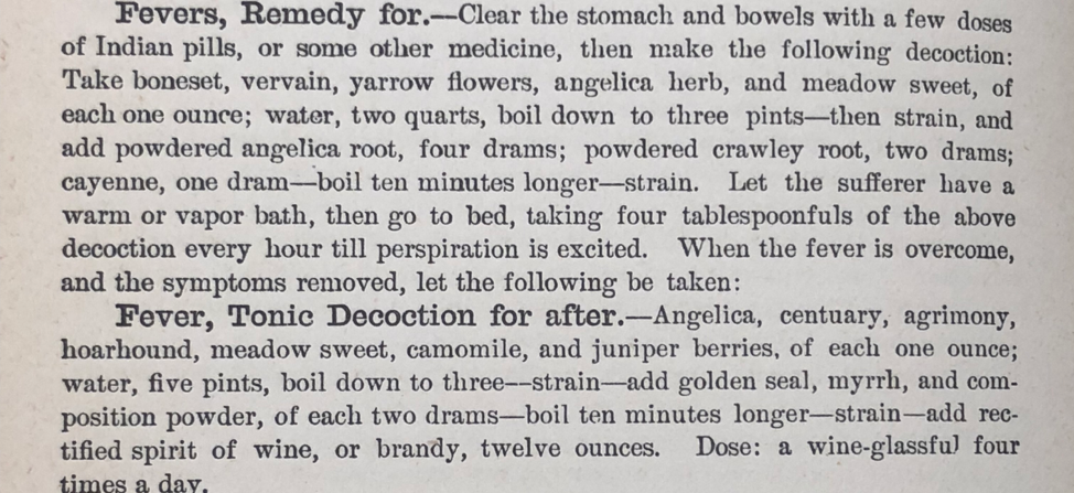
Cottage Physician Treatment for Fever


Ep-PAINE-nym


Kocher Incision


Other Known Aliasesright subcostal incision


Definitionoblique incision in the right subcostal area starting 2-5cm below the xiphoid process, running parallel to the ribs, and extending to 2.5cm below the inferior costal margins


Image result for kocher incision


Clinical Significanceclassic incision used for open cholecystectomy


Kocher Maneuver


Other Known Aliasesretroperitoneal exposure


Definition – maneuver to expose the retroperitoneal structure (pancreas, duodenum, vena cava)


Clinical Significancethis maneuver is used for resection of pancreatic head tumors or in trauma with retroperitoneal hemorrhage.  The peritoneum is incised at the right edge of the duodenum and continued in a “C” shaped from superior to inferior.  The duodenum and head of the pancreas is then reflected medially to the left to expose the retroperitoneal structures.

Image result for kocher maneuver exposure

History – Named after Emil Theodor Kocher (1841-1917), a Swiss surgeon who obtained his medical doctorate from the University of Bern in 1865.  He was arguably the most accomplished surgeon of his time and true pioneer in the field of surgery by promoting and advocating the use of aseptic technique, meticulous dissection with attention to minimal blood loss, and implementation of the scientific method in surgery. His “Text-Book of Operative Surgery” was the definitive guide to surgery in the early 1900s.  He received the Nobel Prize in Physiology and Medicine in 1909…the first Swiss citizen and first surgeon to do so.  The practice of modern surgery would certainly not be where it is today without the work of Dr. Kocher and his other notable eponyms are:


  • Kocher forceps – hemostatic tooth and groove forceps
  • Kocher’s point – entry point for intrventricular catheter to drain CSF from the cerebral ventricles
  • Kocher-Debre-Semelaigne Syndrome – hypothyroidism in infancy
  • Kocher collar incision – used in thyroid surgery
  • Kocher’s sign – eyelid phenomenon in hyperthyroidism


Emil Theodor Kocher.jpg


References

  1. Firkin BG and Whitwirth JA.  Dictionary of Medical Eponyms. 2nd ed.  New York, NY; Parthenon Publishing Group. 1996.
  2. Bartolucci S, Forbis P.  Stedman’s Medical Eponyms.  2nd ed.  Baltimore, MD; LWW.  2005.
  3. Yee AJ, Pfiffner P. (2012).  Medical Eponyms (Version 1.4.2) [Mobile Application Software].  Retrieved http://itunes.apple.com.
  4. Whonamedit – dictionary of medical eponyms. http://www.whonamedit.com
  5. Up To Date. www.uptodate.com
  6. Kocher T.  Text-Book of Operative Surgery. 1911. https://archive.org/details/textbookofoperat01kochuoft
  7. Gautschi OP, Hildebrandt G. Emil Theodor Kocher (25/8/1841-27/7/1917)–A Swiss (neuro-)surgeon and Nobel Prize winner. British journal of neurosurgery. 2009; 23(3):234-6. [pubmed]
  8. Biography of Theodor Kocher by Nobel Prize Society. https://www.nobelprize.org/nobel_prizes/medicine/laureates/1909/kocher-bio.html

Ep-PAINE-nym


Weitlaner (VIGHT-lahn-er) Retractor


Other Known Aliases – Wheatlander, Wheaty


DefinitionSelf-retaining retractor

Image result for weitlaner retractor

Clinical SignificanceOne of the more common self-retaining retractors used in surgery.  They have a ratchet locking system and may also be available with jointed hinges.  Primarily used for small to medium incision exposures.

History – Named after Franz Weitlaner (1872-1944), an Austrian physician who received his medical doctorate from Innsbruck Medical University in Austria at the age of 26 in 1898.  He enjoyed a prolific career as as ship surgeon and house physician practicing in St. Poelten and Ottenthal in his homeland.  In 1905, he published an article in the Vienna Clinical Review entitled “Ein Automatischer Wundspreizer” (An Automatic Wound-spreader), which would be the first description of his famous retractor.  They were originally manufactured by Windler Instrument Makers in Berlin in 1912, but Weitlaner never patented his design or received any monetary gains from his invention, only the right to have the instrument named after him.


References

  1. Firkin BG and Whitwirth JA.  Dictionary of Medical Eponyms. 2nd ed.  New York, NY; Parthenon Publishing Group. 1996.
  2. Bartolucci S, Forbis P.  Stedman’s Medical Eponyms.  2nd ed.  Baltimore, MD; LWW.  2005.
  3. Yee AJ, Pfiffner P. (2012).  Medical Eponyms (Version 1.4.2) [Mobile Application Software].  Retrieved http://itunes.apple.com.
  4. Whonamedit – dictionary of medical eponyms. http://www.whonamedit.com
  5. Up To Date. www.uptodate.com
  6. Sharma A, Swan KG. Franz Weitlaner: the great spreader of surgery. The Journal of Trauma. 2009; 67(6):1431-4. [pubmed]
  7. Weitlaner F. Ein automatischer Wundspreizer. Wien klin Rundschau. 1905;xix:114.

Ep-PAINE-nym


Varess Needle


Other Known AliasesNone

Definition12-15cm long, 2 cannula instrument used for insuflating the abdominal cavity before laparoscopic port placement.  The outer cannula has a beveled needle for dissecting through the abdominal wall.  The spring-loaded inner stylet resides within the outer cannula and has a dull tip to prevent injury to abdominal viscera.  Due to this spring-loaded mechanism, the inner stylet retracts into the outer cannula while it moves through the abdominal planes and advances past the sharp, cutting tip of the outer cannula once through the peritoneum.

Image result for veress needleImage result for veress needle

Clinical Significance Using the Varess needle is the oldest and most traditional techniques for obtaining laparoscopic access

History – Named after János Vares (1903-1979), a Hungarian internist, who used iatrogenic pneumothoraces to treat tuberculosis patients.  He created this spring loaded needle in 1932 and published his results in 1936 (in a Hungarian journal), which was subsequently translated and published in German for wider audience in 1938.  Raoul Palmer (1904-1985), a French gynecologist, began using the Varess needle for laparoscopic surgery in 1947.


References

  1. Firkin BG and Whitwirth JA.  Dictionary of Medical Eponyms. 2nd ed.  New York, NY; Parthenon Publishing Group. 1996.
  2. Bartolucci S, Forbis P.  Stedman’s Medical Eponyms.  2nd ed.  Baltimore, MD; LWW.  2005.
  3. Yee AJ, Pfiffner P. (2012).  Medical Eponyms (Version 1.4.2) [Mobile Application Software].  Retrieved http://itunes.apple.com.
  4. Whonamedit – dictionary of medical eponyms. http://www.whonamedit.com/
  5. Vares J. Neues instrument zur ausfuhrung von brust-oder bauchpunktionen und pneumothoraxbehandlung. Deut Med Wochenschr. 1938;64:1480-1481.
  6. Palmer R. Instrumentation et technique de la coelioscopie gynécologique. Gynecologie et obstetrique. 1947; 46(4):420-31. [pubmed]

PAINE #PANCE Pearl – Surgery


Question


  1. What are the five classic causes of a post-operative fever?
  2. What are two other clever causes to think of (if I do say so myself)?


Answer


  1. The five classic causes of post-operative fever are:
    1. Wind = lungs (atelectasis, pneumonia, aspiration) = POD 1-2
    2. Water = UTI = POD 2-3
    3. Wound = surgical site infection = POD 3-5
    4. Walking = DVT = POD 3-5
    5. Wonder drugs = drug reactions = anytime
  2. Another two “W’s” to add to this list:
    1. Withdrawal = typically alcohol
    2. “Wonky” glands = thyrotoxicosis, adrenal crisis


Check out my article in JAAPA from 2016 on “Evaluating Postoperative Fever” for a more in-depth look

Ep-PAINE-nym


Glisson’s Capsule


Other Known AliasesNone

DefinitionOuter capsule of connective fibrous tissue, surrounding the liver, the intrahepatic branches of the portal vein, hepatic arteries, and bile duct

Clinical Significance The is a structure that must be dissected while operating on the liver.  In trauma, you can have subcapsular hematomas from hemorrhage that are contained by Glisson’s capsule.

History – Named after Francis Glisson (1597-1667), who was an English physician, anatomist, and pathologist.  His work on the liver in the late 1600s produced the foremost textbook on the digestive system, The Anatomia Hepatis, where he first described the covering of the liver in detail.


References

  1. Firkin BG and Whitwirth JA.  Dictionary of Medical Eponyms. 2nd ed.  New York, NY; Parthenon Publishing Group. 1996.
  2. Bartolucci S, Forbis P.  Stedman’s Medical Eponyms.  2nd ed.  Baltimore, MD; LWW.  2005.
  3. Yee AJ, Pfiffner P. (2012).  Medical Eponyms (Version 1.4.2) [Mobile Application Software].  Retrieved http://itunes.apple.com.
  4. Whonamedit – dictionary of medical eponyms. http://www.whonamedit.com/
  5. Haubrich WS. Glisson of Glisson’s capsule of the liver. Gastroenterology. 2001; 120(6):1362. [pubmed]

#31 – Small Bowel Obstruction


***LISTEN TO THE PODCAST HERE***


Epidemiology

Small bowel obstruction (SBO) is one of the more common surgical emergencies in US, due to the increased intraluminal pressures which can lead to intestinal ischemia and risk of spontaneous rupture.  Overall mortality increases significantly if either of these occur.  It is estimated that over 300,000 surgeries per year occur in the US for bowel obstructions, with the small bowel making up around 80%.  Although it can happen at age, there is a higher trend to older patients and thee is equal incidence with both genders.


Risk Factors

The most common cause of SBO is postoperative adhesions, but there are numerous other causes that you should keep on your differential.


Signs and Symptoms

  • Abdominal pain
    • Paroxysms of periumbilical pain (3-5 minutes)
  • Nausea
  • Vomiting
  • Obstipation

Physical Examination

  • Dehydration
    • Tachycardia, orthostasis, decreased urine output
  • +/- Distension
  • Changes in bowel sounds
    • Hyperactive à muffled à absent
  • Percussion changes
    • Tympanic with distension
    • Dullness with fluid
  • Peritoneal signs if perforation present
  • +/- hemoccult

Laboratory Studies

  • CBC with differential
    • Anemia can point to a chronic condition
  • BMP
  • ABG
    • Acidosis = bowel ischemia, volume depletion
    • Alkalosis = vomiting
  • Serum lactate

Imaging Studies

  • Plain Radiographs
    • Dilated loops of small bowel with air/fluid levels on upright film
    • May also see a paucity of gas in distal intestines
    • Small bowel dilation ≥ 2.5cm is diagnostic

  • Abdominal CT
    • Much more sensitive and can also identify the specific causes
    • Can also identify a transition point
    • Other radiological signs on CT consistent with SBO include:
      • Bowel wall thickening > 3mm
      • Submucosal edema
      • Mesenteric edema
      • Ascites
      • Target sign (intussusception)
      • Whirl sign (volvulus)
      • Venous cut-off sign (thrombosis)
  • Ultrasound
    • Not as good as CT, but better than plain radiography
  • Special Considerations on Radiography
    • Closed-Loop Obstruction
      • High risk for ischemia, perforation, and ischemic bowel
    • Ischemia and Perforation

Management

  • Initial
    • NPO
    • Fluid resuscitation
    • Surgery consultation
    • Gastrointestinal decompression
      • Not in EVERY patient, but good for patients with significant distension, nausea, and/or vomiting
    • Surgery
      • Nonspecific signs of bowel ischemia:
        • Fever
        • Leukocytosis
        • Tachycardia
        • Continuous and/or worsening abdominal pain
        • Metabolic acidosis
        • Peritonitis
      • High likelihood of bowel resection if ≥ 3 of the following:
        • Pain > 4 days
        • Abdominal guarding on exam
        • Elevated CRP > 75 mg/dL
        • Leukocytosis > 10,000
        • > 500cc fluid of intraabdominal fluid
        • Reduced wall contrast enhancement on CT
    • Observation
      • If no high-risk surgical signs present, observation with serial examinations may be used for 12-24 hours in patients with:
        • Early postoperative obstruction
        • Inflammatory bowel disease
        • Gallstone ileus
        • Infectious small bowel disease
        • Colonic diverticular disease
      • Exploration should proceed if no improvement

References

  • Miller G, Boman J, Shrier I, Gordon PH. Etiology of small bowel obstruction. American Journal of Surgery. 2000; 180(1):33-6. [pubmed]
  • Markogiannakis H, Messaris E, Dardamanis D. Acute mechanical bowel obstruction: clinical presentation, etiology, management and outcome. World Journal of Gastroenterology. 2007; 13(3):432-7. [pubmed]
  • Scott FI, Osterman MT, Mahmoud NN, Lewis JD. Secular trends in small-bowel obstruction and adhesiolysis in the United States: 1988-2007. American Journal of Surgery. 2012; 204(3):315-20. [pubmed]
  • Drożdż W, Budzyński P. Change in mechanical bowel obstruction demographic and etiological patterns during the past century: observations from one health care institution. Archives of Surgery (Chicago, Ill. : 1960). 2012; 147(2):175-80. [pubmed]
  • Taylor MR, Lalani N. Adult small bowel obstruction. Academic Emergency Medicine. 2013; 20(6):528-44. [pubmed]
  • Jackson PG, Raiji MT. Evaluation and management of intestinal obstruction. American Family Physician. 2011; 83(2):159-65. [pubmed]
  • Mullan CP, Siewert B, Eisenberg RL. Small bowel obstruction. American Journal of Roentgenology. 2012; 198(2):W105-17. [pubmed]
  • Suri S, Gupta S, Sudhakar PJ, Venkataramu NK, Sood B, Wig JD. Comparative evaluation of plain films, ultrasound and CT in the diagnosis of intestinal obstruction. Acta radiologica. 1999; 40(4):422-8. [pubmed]
  • Zalcman M, Sy M, Donckier V, Closset J, Gansbeke DV. Helical CT signs in the diagnosis of intestinal ischemia in small-bowel obstruction. American Journal of Roentgenology. 2000; 175(6):1601-7. [pubmed]
  • Eltarawy IG, Etman YM, Zenati M, Simmons RL, Rosengart MR. Acute mesenteric ischemia: the importance of early surgical consultation. The American Surgeon. 2009; 75(3):212-9. [pubmed]
  • Schwenter F, Poletti PA, Platon A, Perneger T, Morel P, Gervaz P. Clinicoradiological score for predicting the risk of strangulated small bowel obstruction. The British Journal of Surgery. 2010; 97(7):1119-25. [pubmed]