What is a good mnemonic for the red flag history and/or symptoms of acute back pain?
Answer
The majority of the back pain you will see in clinical practice is non-emergent, but you need to be able to identify the cases that need emergent referral, consultation, or imaging. Just remember TUNAFISH……
Trauma
Any trauma can cause fracture and cord compromise. Back pain + trauma = imaging
Unintentional weight loss
Think vertebral metastasis of cancer
Neurologic deficits
Big ones are saddle anesthesia and bowel/bladder dysfunction. Paresthesias, weakness, and numbness are concerning, but emergent.
Age > 50
New-onset back pain in patients > 50 years old can be cancer, infection, or AAA
Fever
Again…think osteomyelitis, spinal abscess, or cancer
IVDU
Hematogenous infectious seeding of the vertebral bodies or spinal abscess
Steroid Use
Chronic steroid use weakens bones and even low energy mechanisms or spontaneous fractures are possible
History of cancer
Metastases
References
Della-Giustina D. Evaluation and treatment of acute back pain in the emergency department. Emergency medicine clinics of North America. 2015; 33(2):311-26. [pubmed]
Borczuk P. An evidence-based approach to the evaluation and treatment of low back pain in the emergency department. Emergency medicine practice. 2013; 15(7):1-23; Quiz 23-4. [pubmed]
Other Known Aliases – Tarsometatarsal fracture/dislocation
Definition – Fracture/dislocation of the articulation of the tarsal bones with the metatarsals of the foot.
Clinical Significance – The Lisfranc joint of the foot is where the first three metatarsals articulate with the three cuneiforms and the fourth and fifth metatarsals articulate with the cuboid. The Lisfranc ligament attaches the medial cuneiform to the 2nd metatarsal bone on the the plantar surface of the foot. This is a very serious injury of the foot and sometimes may simple present as a bad sprain. This injury is most common seen with direct crush injuries and indirect load onto a plantar flexed foot.
History – This injury was first described by Jacques Lisfranc de St. Martin (1790-1847), a French surgeon who served in Napoleon’s army in 1813. He noted this injury pattern in Calvary soldiers who fell from their horse and caught their foot in the stirrup.
References
Firkin BG and Whitwirth JA. Dictionary of Medical Eponyms. 2nd ed. New York, NY; Parthenon Publishing Group. 1996.
Bartolucci S, Forbis P. Stedman’s Medical Eponyms. 2nd ed. Baltimore, MD; LWW. 2005.
Yee AJ, Pfiffner P. (2012). Medical Eponyms (Version 1.4.2) [Mobile Application Software]. Retrieved http://itunes.apple.com.
Definition – Any fracture dislocation of the talus.
Clinical Significance – None. This is an antiquated term for talar injuries
History – First coined in 1919 by Henry Graeme Anderson, who was a consulting surgeon for the Royal Flying Corps during World War I. He described 18 cases of fracture and dislocation of the talus in pilots between 1914-1919. During the early history of flight, planes did not reach lethal speeds and when they crashed, the rudder bar (which was controlled by the pilot’s feet) would get driven up into the instep of the foot just anterior to the calcaneous.
References
Firkin BG and Whitwirth JA. Dictionary of Medical Eponyms. 2nd ed. New York, NY; Parthenon Publishing Group. 1996.
Bartolucci S, Forbis P. Stedman’s Medical Eponyms. 2nd ed. Baltimore, MD; LWW. 2005.
Yee AJ, Pfiffner P. (2012). Medical Eponyms (Version 1.4.2) [Mobile Application Software]. Retrieved http://itunes.apple.com.
Other Known Aliases – Sound-induced vestibular activation.
Definition – Vertigo, dizziness, nausea, and nystagmus caused by a load noise.
Clinical Significance – This pathology is due to a communication between the middle and inner ear classically associated with congenital syphilis. Recently, it has been associated with superior canal dehiscence syndrome (SCDS). This can also be elicited with nose-blowing, valsalva, and heavy lifting.
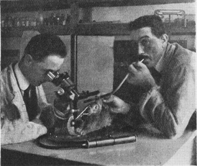
History – Named after Italian biologist Pietro Tullio, Ph.D. (1881-1941), who originally studied this finding in pigeons and published it in 1929.
Tullio blowing a whistle in the ear of rabbit test subject
References
Firkin BG and Whitwirth JA. Dictionary of Medical Eponyms. 2nd ed. New York, NY; Parthenon Publishing Group. 1996.
Bartolucci S, Forbis P. Stedman’s Medical Eponyms. 2nd ed. Baltimore, MD; LWW. 2005.
Yee AJ, Pfiffner P. (2012). Medical Eponyms (Version 1.4.2) [Mobile Application Software]. Retrieved http://itunes.apple.com.
Definition – Small, bilateral pupils with an absence of miotic reaction to light, both direct and consensual, with preservation of miotic reaction to near stimulus. In other words, they accommodate, but do not react light (light-near dissociation).
Clinical Significance – Classically associated with tabes dorsalis of neurosyphylis, but can also be seen in diabetic neuropathy. Rare now due to the widespread of antibiotics and treating early syphilis infections
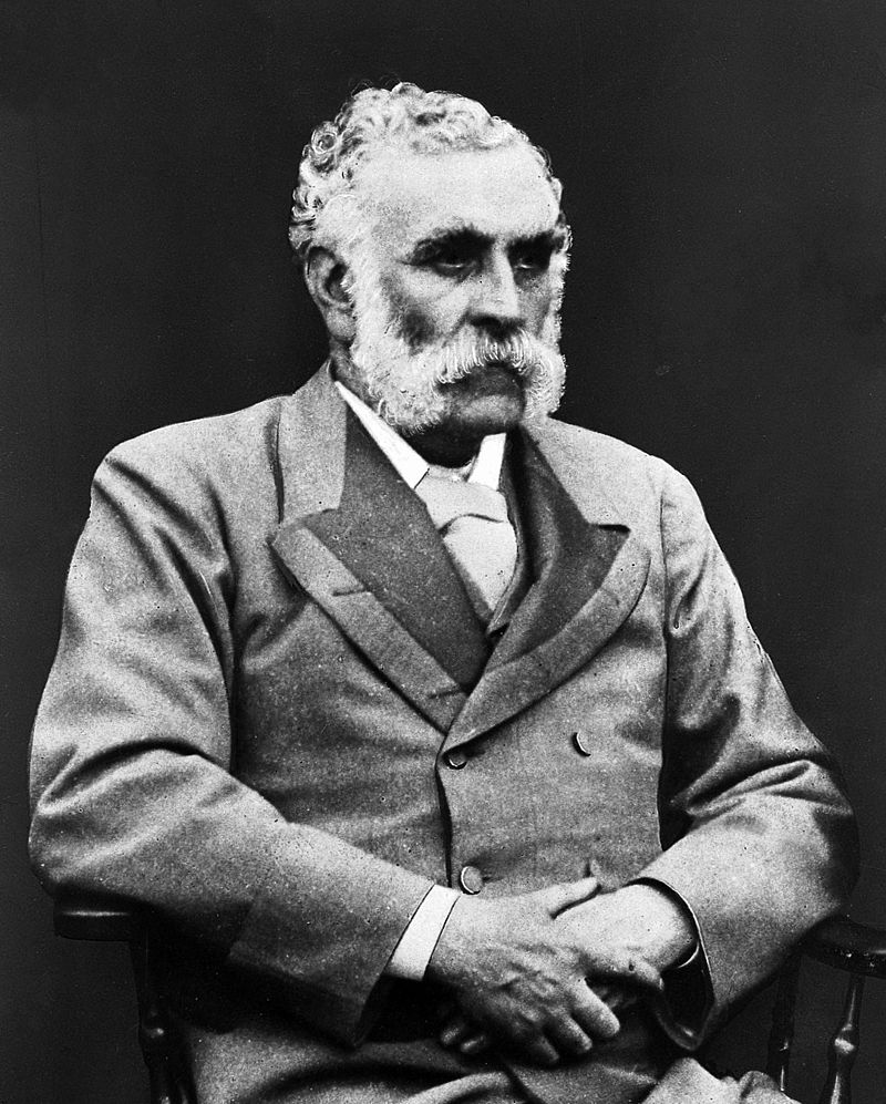
History – Named after Douglas Moray Cooper Lamb Argyll Robertson (1837-1909), who was a Scottish surgeon and ophthalmologist and one of the first to specialize in the eye. He published his findings of several case reports in two articles in the “Edinburgh Medical Journal” in 1869. Previous to this however, he was also the first to discover and use the extract of the Calabar bean (otherwise known as physostigmine) for treatment of various eye disorders.
“Dougie”, as his friends called him****
References
Firkin BG and Whitwirth JA. Dictionary of Medical Eponyms. 2nd ed. New York, NY; Parthenon Publishing Group. 1996.
Bartolucci S, Forbis P. Stedman’s Medical Eponyms. 2nd ed. Baltimore, MD; LWW. 2005.
Yee AJ, Pfiffner P. (2012). Medical Eponyms (Version 1.4.2) [Mobile Application Software]. Retrieved http://itunes.apple.com.
Robertson DA. On an interesting series of eye symptoms in a case of spinal disease, with remarks on the action of belladonna on the iris. Edinb Med J. 1869;14:696–708.
Robertson DA. Four cases of spinal myosis with remarks on the action of light on the pupil. Edinb Med J. 1869;15:487–493
Robertson, D. A.: On the Calabar Bean as a New Agent in Ophthalmic Medicine. Edinb Med J. 1863;93:815-820.
****I have no source for this but he looks like a Dougie….plus with a name like Douglas Moray Cooper Lamb Argyll Robertson, you have to have a nickname, right?
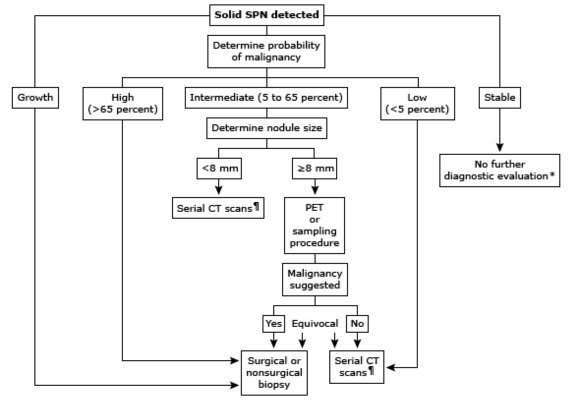
When following low-risk, small single pulmonary nodules,
What is the time sequence for the follow-up CT scans?
How long do you re-image them for?
Answers
It depends on the size of the nodule and consistency after the initial CT scan. The current guidelines state:
For solid nodules:
< 6mm recommend no further imaging
> 6mm but < 8mm recommend CT scan at 6-12 months
> 8mm recommend CT scan at 3-6 months, 9-12 months, and 18-24 months
For non-solid (ground glass) nodules:
< 5mm recommend no further imaging
> 5mm recommend annual CT for 3 years
For part-solid (>50% ground glass) nodules:
< 8mm recommend CT scan at 3 months, 12 months, 24 months, and annual for 1-3 years
> 8mm recommend CT scan at 3 months and PET or biopsy
Low-risk nodules can be serially scanned for 2 years and if no change, can stop repeat imaging as malignant nodules will show some form of change in 2 years.
References
Gould MK, Donington J, Lynch WR. Evaluation of individuals with pulmonary nodules: when is it lung cancer? Diagnosis and management of lung cancer, 3rd ed: American College of Chest Physicians evidence-based clinical practice guidelines. Chest. 2013; 143(5 Suppl):e93S-e120S. [pubmed]
300,000-600,000 cases per year in the United States
It is estimated that up to 50% will have post-thrombotic syndrome
Why Are We So Scared?
As many as 20% of patients with 1st onset PTE have no identifiable risk factors
10-30% 1-month mortality with up to 25% presenting as sudden death
Fear of litigation is #1 reason clinicians work-up low risk PTE
Why Can’t We Test Everyone?
Up to $16,000 per patient in total health care costs
6 times more deaths with testing and treatment
Signs and Symptoms
The majority of the classic signs and symptoms come from PIOPED II study and EMPEROR registry. These include:
Dyspnea (73%)
Chest Pain (64%)
Tachypnea (57%)
DVT findings or leg pain/swelling (47%)
Tachycardia (26%)
Dizziness (12%)
Hemoptysis (10%)
The EMPEROR registry took it a step further and determined mean vital sign measurements of:
Heart rate – 95 bpm
Respiratory rate – 20 bpm
Oxygen saturation – 95%
A recent trail in the NEJM called PESIT concluded as many as 1 in 6 patients with first time syncope has a PTE on inpatient work-up. This study has been largely panned by the EM community and you can read their take from the links below:
The most well know score is the Wells Criteria first published in 1998 and then revised and simplified in 2000 and 2001.
A second calculation is the Geneva Score first published in 2001 and revised and simplified in 2006 and 2008.
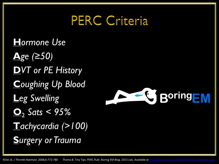
The Pulmonary Embolism Rule-Out Criteria was published in 2008 by Jeff Kline and is a second set of criteria to definitively rule-out PTE in patients ALREADY SCORE AS LOW RISK by Wells or Geneva.
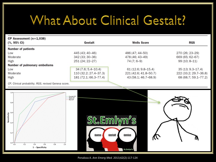
What about just good ol’ clinical gestalt? An interesting study was performed in 2013 looking at the accuracy of Wells vs Geneva vs Gestalt and found:
Clinical gestalt had a lower missed rate of PTE in low-risk patients
Clinical gestalt had a high accuracy of diagnosing PTE in high-risk patients
The Work-Up of Suspected PTE
Electrocardiogram is not senstitive nor specific for PTE but should be ordered on every patient with chest pain and/or shortness of breath to rule-out ACS
The EMCMD talks about the 10 ECG findings of PTE in the best video I have every scene
D-Dimer
High senstivity = good for rule-out
Should only be used after pre-test probability due to the false positives and unnecessary work-ups
ADJUST-PE Study
Found D-Dimer go up with age and created an age adjusted D-Dimer cutoff of:
Age (yr) x 10 as diagnostic threshold
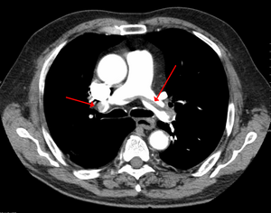
Radiographic Imaging
Computed tomography is gold standard but has higher radiation exposure and contrast loads
Ventilation/Perfusion scan is safer in renal patients but up to 2/3rd are non-diagnostic
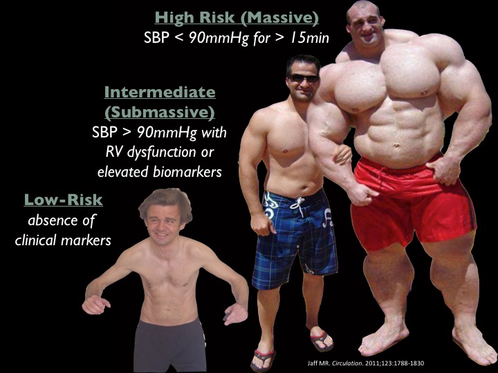
Risk Assessment
Once you diagnose a patient with a PTE, you have determine the patient’s risk and severity of disease.
Echocardiogram
Looking for RV strain
RV:LV ≥ 1
RV hypokinesis
Paradoxical septal movement
Tricuspid regurgitation
Biomarkers
Brain Natriuretic Peptide (BNP)
> 90 pg/mL has been associated with increased mortality
Troponin
> 0.01 ng/mL suggests evidence of RV dysfunction
Pulmonary Embolism Severity Index (PESI)
Published in 2005 and simplified 2010
Developed to help prognosticate 30d mortality and found low-risk patients (PESI – 0) can be safely treated as outpatient
Definitions/Grades of PTE
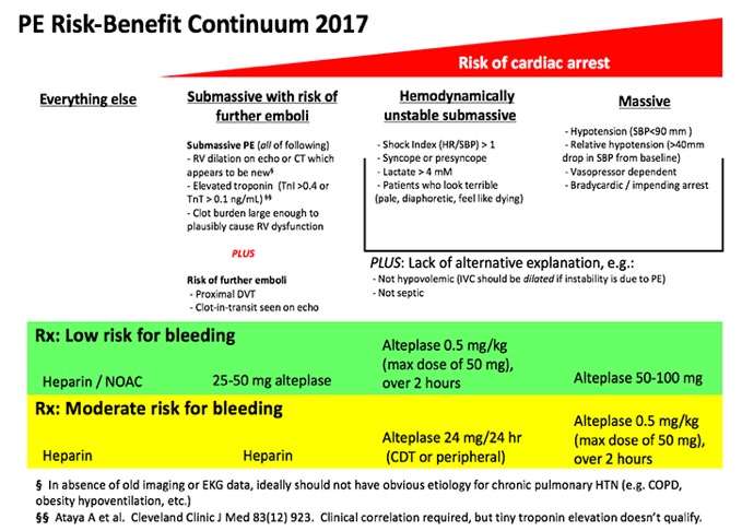
Treatment Strategies for PTE
Anticoagulation
Started with confirmation of PTE or with high pre-test probability during workup
Lots of options (heparin, LMWH, direct thrombin inhibitors, Factor Xa inhibitors)
Fibrinolytics
Lots of recent research on who to lyse and who not to
Original research showed benefit if full dose lytics were given to massive PTE, but harm in submassive patients
This led to MOPPET in 2013 evaluating 1/2 dose lytics in submassive patients and found:
Reduction in overall mortality
No difference in bleeding complications
Reduction in hospital stay
PEITHO came next in 2014 and looked at full dose lytics vs anticoagulation only for submassive PTE and found:
No mortality benefit
Reduction in hemodynamic compromise
Increase in major bleeding and intracranial hemorrhage
Catheter Directed Therapy
Good options in patients with a high risk of bleeding with systemic fibrinolytic therapy
ULTIMA and SEATTLE-II studies found reduction in RV:LV ratio and decreased bleeding complications
Surgical Thrombectomy
Can be used as a last resort option and mortality from these procedures has dramatically improved from 57% in the 1960s to < 6% in 2005
Putting It All Together
This is a graphic I modified from Jeff Kline and EmCrit that encompasses everything into a nice, neat package and as I have said before “algorithms will set you free”
References
Beckman MG. Venous Thromboembolism: A Public Health Concern. Am J Prev Med. 2010;38(4S):S495-S501
Spyropoulous AC. Direct medical costs of venous thromboembolism and subsequent hospital readmission rates: an administrative claims analysis from 30 managed care organizations. J Manag Care Pharm. 2007;13(6):475-486
Calder KK. The mortality of untreated pulmonary embolism in emergency department patients. Ann Emerg Med. 2005;45(3):302-310
Stein PD. Silent pulmonary embolism in patients with deep venous thrombosis: A systematic review. Am J Med. 2010;123:426-431
Stein PD. Clinical Characteristics of patients with acute pulmonary embolism. Am J Med. 2007;120:871-879
Pollack CV. Clinical characteristics, management, and outcoms of patients diagnosed with acute pulmonary embolism in the emergency department. JACC. 2011;57(6):700-706
Wells PS. Use of a clinical model for safe management of patients with suspected pulmonary embolism. Ann Intern Med. 1998;129:997-1005
Wells PS. Derivation of a simple clinical model to categorize patients probability of pulmonary embolism: increasing the models utility with the SimpliRED D-dimer. Thromb Haemost. 2000;83:416-420
Wells PS. Excluding pulmonary embolism at the bedside without diagbnostic imaging: management of patients with suspected pulmonary embolism presenting to the emergency department by using a simple clinical model and D-dimer. Ann Intern Med. 2001;135:98-107
van Belle A. Effectiveness of managing suspected pulmonary embolism using an algorithm combining clinical probability, D-dimer testing, and computed tomography. JAMA. 2006;295(2):172-179
Wicki J. Assessing clinical probability of pulmonary embolism in the emergency ward: a simple score. Arch Intern Med. 2001;161:997-92-97
Le Gal G. Prediction of pulmonary embolism in the emergency department: the revised Geneve score. Ann Intern Med. 2006;144:165-171
Klok FA. Simplication fo the revised Geneva score for assessing clinical probability of pulmonary embolism. Ann Intern Med. 2008;168(19):2131-2136
Kline JA. Prospective multicenter evaluation of the pulmonary embolism rule-out criteria. J Thromb Haemost. 2008;6:772-780
Penaloza A. Comparison of the unstructured clinical gestalt, the wells score, and the revised Geneva score to estimate pretest probability for suspected pulmonary embolism. Ann Emerg Med. 2013;62(2):117-124
Righini M. Age-adjusted D-dimer cutoff levels to rule-out pulmonary embolism: the ADJUST-PE study. 2014;311(11):1117-1124
Stein PD. Clinical characteristics of patients with acute pulmonary embolism: data from IOPED II. Am J Med. 2007;120:871-879
Anderson DR. Computerized tomographic pulmonary angiography versus ventilation perfusion lung scanning for the diagnosis of pulmonary embolism. Curr Opin Pulm Med. 2009;15:425–429
Rudoni RR. Use of two-dimensional echocardiography for the diagnosi of pulmonary embolus. J Emerg Med. 1998;16(1):5-8
Taylor RA. Point-of-care focused cardiac ultrasound for prediction of pulmonary embolism adverse outcomes. J Emerg Med. 2013;45(3):392-399
Kiely DG. Elevated levels of natriuretic peptides in patients with pulmonary thromboembolism. Resp Med. 2005;99:1286-1291
Jaff MR. Management of massive and submassive pulmonary embolism, iliofemoral deep vein thrombosis, chronic thromboembolic pulmonary hypertension: a scientific statement form the American Heart Association. Circulation. 2011;123:1788-1830
Keller K. Cardiac troponin I for predicting right ventricular dysfunction and intermediate risk in patients with normotensive pulmonary embolism. Neth Heart J. 2015;23:55-61
Aujesky D. Derivation and validation of a prognostic model for pulmonary embolism. Am J Respir Crit Care Med. 2005;172:1041-1046
Jimenez D. Simplification of the pulmonary embolism severity index for prognostication in patients with acute symptomatic pulmonary embolism. Arch Intern Med. 2010;170(15):1383-1389
Tapson VF. Treatment of pulmonary embolism: anticoagnulation, thrmbolytic therapy, and complications of therapy. Crit Care Clin. 2011;27:825-839
Sharifi M. Moderate pulmonary embolism treated with thrombolysis. Am J Cardiol. 2013;111:273-277
Zhang Z. Lower dosage of recombinant tissue-type plasminogen activator (rt-PA) in the treatment of acute pulmonary embolism: a systematic review and meta-analysis. Thrombosis Research. 2014;133:357-363
Meyer GM. Fibrinolysis for patients with intermediate-risk pulmonary embolism. NEJM. 2014;370(15):1402-1411
Chatterjee S. Thrombolysis for pulmonary embolism and risk of all-cause mortality, major bleeding, and intracranial hemorrhage: a meta-analysis. JAMA. 2014;311(23):2414-2421
Curtis GM. Risk factors associated with bleeding after alteplase administration for pulmonary embolism: a case control study. 2014;34(8):818–825
Kennedy RJ. Thrombus resolution and hemodynamic recovery using ultrasound-accelerated thrombolysis in acute pulmonary embolism. J Vasc Interv Radiol. 2013;24:841-848
Kucher N. Randomized, controlled trial of ultrasound-assisted catheter-directed thrombolysis for acute intermediate-risk pulmonary pulmonary embolism. Circulation. 2014;129:479-486
Cross FS. A survey of the current status of pulmonary embolectomy for massive pulmonary embolism. Circulation. 1967;35:186-191
Stulz P. Decision making in the surgical treatment of massive pulmonary embolism. Eur J Cardio-thorac Surg. 1994;8:188-193
Leacche M. Modern surgical treatment of massive pulmonary embolism: result in 47 sonsecutive patients after rapid diagnosis and aggressive surgical approach. J Thorac Cardiovasc Surg. 2005;129:1018-1023
Prandoni P, et al (PESIT Investigators). Prevalance of Pulmonary Embolism amount Patients Hospitalized for Syncope. NEJM. 2016;375:1524-1531
John MA, Klok FA, van Es N. D-dimer Interval Likelihood Ratios for Pulmonary Embolism. Acad Emerg Med. 2017;4;1-5.
Konstantinides SV, et al. Impact of Thrombolytic Therapy on the Long-Term Outcome of Intermediate-Risk Pulmonary Embolism. JACC. 2017;69(12):1536-1544
Sharifi M, et al (PEAPETT Investigators). Pulseless electrical activity in pulmonary embolism treated with thrombolysis. Am J Emerg Med. 2016;34(10):1963-1967
Piazza G, et al. (SEATTLE-II Investigators). A prospective, single arm, multicenter trial of ultrasound-facilitated , catheter-directed, low-dose fibrinolysis for acute massive and submassive pulmonary embolism. JACC. 2015;8(1):1382-1392