OB/GYN Case #2

27yo G1P1001 presents to the gynecologist’s office to get established as a new patient and have her annual well woman examination performed.  She has had no GYN complaints over the past year and is not currently on her cycle.

Past Medical History

None

Medications

Ortho Tri-Cyclen

Past Obstetric History

38-week NSVD without complications

Past Gynecologic History

Age of menarch – 13

Coital debut – 17

Lifetime partners – 4

Cycles regular at 28-30 days and lasting for 3-5 days with mild/moderate bleeding

Last pap – 2014 and normal per patient No history of STI

Vital Signs

BP-124/75, HR-74, RR-14, O2-100%, Temp-99.1

Physical Exam

Abdomen – Soft, non-tender, non-distended

GU – no cervical motion tenderness, no adenexal tenderness

healthy-cercix

Pap Results

Atypical squamous cells of undetermined significance


Answer will be posted on 02/20


Answer to OB/GYN Case #1

Answers

Diagnosis: placenta previa

Management: ultrasound

Risk factors: hypertension is NOT a risk factor

Discussion

Placenta previa is a condition where the placenta has attached over the cervical os and can be complete, partial, marginal, or low-lying depending on the position.

previa

The hallmark of placenta previa is painless, 2nd trimester bleeding.  Ultrasound should always be performed prior to any speculum or digital exam to limit the risk of bleeding.

Risk factors for placenta previa include :

Previous placenta previa

Previous cesarean delivery (risk increases with an increasing number of cesarean deliveries)

Multiple gestation

Multiparity

Advanced maternal age

Infertility treatment

Previous abortion

Previous intrauterine surgical procedure

Maternal smoking

Maternal cocaine use

Male fetus

Non-white race

OB/GYN Case #1

30-year-old, 32 week gravid,  G5 P3013 female presents to clinic with vaginal bleeding.  She denies any contractions, leakage of fluid, or trauma.  She states that 4 weeks previously she engaged in vaginal intercourse with her husband and had some mild “spotting” that spontaneously resolved over the course of 24 hours.  BP-110/60, HR-97, RR-20, Temp-98.9o F, and O2 saturation 100% on room air.  Physical examination reveals a soft, non-tender abdomen with fundal height approximately 30cm from pubic symphasis.  Fetal heart tones are present and range from 140-150 bpm.

I will post the answer in 1 week.

#7 – Zika Virus

What is it?

The Zika virus is a flavivirus that is related to yellow fever, dengue fever, West Nile, and Japanese encephalitis.  It was first discovered in 1947 in a rhesus monkey and is called “Zika” because it originated in the Zika forest in Uganda.  It is transmitted by the Aedes species of mosquitoes (which also carries dengue, chikungunya, and yellow fever).

Kaddumukasa MA. J of Medical Entomology. 2014;51(1):104-113

Kaddumukasa MA. J of Medical Entomology. 2014;51(1):104-113

Whats with all the noise?

In May 2015, the Pan American Health Organization (PAHO) issued an alert regarding the first confirmed Zika virus infection in Brazil.  Since then, there have documented cases of Zika virus infections in 20 countries in North and South America.  Due to the threat and concern of transmission, the Center for Disease Control (CDC) issued a travel alert (Level 2-Practice Enhanced Precautions) for anyone traveling to these regions (see current list here).

Screen Shot 2016-02-01 at 11.48.58 AM

CDC Travel Alert Notice

Map2

CDC Map of Reported Active Transmission

Map3

McNeil DG. SHort Answers to Hard Questions About Zika. New York Times. 2016

Signs and Symptoms

Only 1 in 5 people who become infected by the Zika virus develop symptoms, which are usually a mild viral prodromal syndrome (fever, rash, arthralgias, myalgias, conjunctivitis).  Serious manifestations can include Guillaine-Barre syndrome and congenital microcephaly.  Brazil has seen a 20-fold increase in number of cases of microcephaly from 2010 to 2014.  Since October 2015, there have been 4,180 suspected cases (average of ~150/yr) of microcephaly in Brazil, but only 700 mothers were tested for the virus with only 270 positive results.  The first case of microcephaly associated by the Zika virus on US soil was in Hawaii on January 15th, 2016 to a mother who lived in Brazil.

Map4

McNeil DG. Short Answers to Hard Questions About Zika. New York Times. 2016.

Because of the surge of microcephaly cases in an endemic region of Zika, the CDC is recommending that women who are pregnant, or are trying to become pregnant, should post-pone any travel to these regions.

Map5

McNeil DG. Short Answers to Hard Questions About Zika. New York Times. 2016.

Testing

Signs and symptoms of the acute Zika infection are very non-specific and the list of differential diagnoses is broad.  If a patient has any suspicious symptoms within one week of travel to any of the at-risk regions, testing should occur as the Zika virus has become a nationally notifiable condition by the CDC.  Testing includes reverse transcriptase-polymerase chain reaction (RT-PCR), virus-specific IgM and neutralizing antibodies and are only performed at the CDC Arbovirus Diagnostic Laboratory.  Clinicians should contact local health departments to facilitate obtaining the correct testing sample and expediting transfer to the lab.

Treatment and Prevention

There is no specific treatment for the Zika virus.  Treatment plans should be directed towards symptom relief and includes rest, oral rehydration, and acetaminophen for fever and pain relief.  Aspirin and other NSAIDs should be avoided until dengue fever can be ruled out to decrease the risk of hemorrhage.

If travel must occur to endemic regions, patients should be advised to follow strict mosquito precautions to try to limit the exposure from the Aedes vector.  N,N-Diethyl-meta-toluamide (DEET), Picaridin, oil of lemon eucalyptus, and IR3535 are all recommended by the CDC and are safe in pregnancy.  There is no vaccine to the Zika virus yet, but preliminary works seem to be promising and early reports are pointing to the end of 2016 as a reasonable estimate for human trials to start.

Repellents

CDC Recommendations for Repellents

Bottom Line

For a PA practicing in the United States, this just adds to the list of traveler’s disease that you have to be hypervigilant about in patients with general complaints and recent travel to endemic regions.  By no means do we need to start screening every patient with viral symptoms for Zika.  But…if your patient has traveled to these regions, is pregnant, or may come into contact with patients who are pregnant, you should contact your local health department and screen them appropriately now that it is a nationally reportable disease.  You should also take appropriate quarantine precautions if your clinic/department also has pregnant patients to limit disease contact to the most at risk patients.  To date, there are no direct transmission cases of the virus (only the Aedes vector), but viruses can shift fast and it is better to be safer than sorry.

References

  1. Lucey DR, Gostin LO. The Emerging Zika Pandemic: Enhancing Preparedness. Published online January 27, 2016. doi:10.1001/jama.2016.0904.
  2. Kaddumukasa MA, Mutebi JP, Lutwama JJ, Masembe C, Akol AM. Mosquitoes of Zika Forest, Uganda: Species Composition and Relative Abundance.  J of Medical Entomology.  2014;51(1):104-113.
  3. (2016, January 29). In Wikipedia, The Free Encyclopedia. Retrieved 17:33, January 31, 2016, from https://en.wikipedia.org/w/index.php?title=Aedes&oldid=702307969
  4. Zika virus. Centers for Disease Control and Prevention Available at: http://www.cdc.gov/zika/.  Accessed February 1, 2016.
  5. Areas with Zika. Centers for Disease Control and Prevention Available at: http://www.cdc.gov/zika/geo/index.html. Accessed February 1, 2016.
  6. Brazil: 270 of 4,180 suspected microcephaly cases confirmed. The Big Story. Available at: http://bigstory.ap.org/article/25bba65de82b437080bedd4f9e229280/brazil-270-4120-suspected-microcephaly-cases-confirmed. Accessed February 1, 2016.
  7. Mcneil DG, Louis CS, St N. Short Answers to Hard Questions About Zika Virus. The New York Times Available at: http://www.nytimes.com/interactive/2016/health/what-is-zika-virus.html. Accessed February 1, 2016.
  8. Diagnostic Testing. Centers for Disease Control and Prevention Available at: http://www.cdc.gov/zika/hc-providers/diagnostic.html. Accessed February 1, 2016.
  9. Could We Have a Zika Vaccine Soon? NBC News. Available at: http://www.nbcnews.com/storyline/zika-virus-outbreak/could-we-have-zika-vaccine-soon-n507186. Accessed February 1, 2016.
  10. The Brazilian Doctors Who Sounded the Alarm on Zika and Microcephaly. WSJ. Available at: http://www.wsj.com/articles/the-brazilian-doctors-who-sounded-alarm-on-zika-and-microcephaly-1454109620?mod=e2tw. Accessed February 1, 2016.


Hypertension Review – JNC-8 and SPRINT

One of the more common conditions that is managed in all of medicine is hypertension.  The first Joint National Committee (JNC) recommendations were published in 1976 and have gone through 8 revisions, with the most recent being in 2014.  The interesting thing about the JNC 8 is that it took 11 years to get published (4-6 years for all the other updates).  Even then there is still some controversy surrounding its recommendations.

 Joint National Committee 8

Emphasis on randomized, controlled clinical trials to answer 3 key clinical questions:

  1. Does initiating antihypertensive pharmacologic therapy at specific blood pressure thresholds improve health outcomes?
    1. When to start
  2. Do attempts to reach specified blood pressure goals with antihypertensive pharmacologic therapy lead to improvements in health outcomes?
    1. What to shoot for
  3. Do various antihypertensive drugs or drug classes differ in regard to specific health outcomes?
    1. What to use

9 Graded Recommendations

  1. Adult patients 60 years of age or older, without specific comorbidities, should have antihypertensive medications initiated if SBP > 150 mmHg or DBP > 90 mmHg and treat to a goal SBP < 150 mmHg and DBP < 90 mmHg.
    1. Corollary
      1. If treated SBP < 140 mmHg is well tolerated and without adverse effects on health or quality of life, no adjustment is needed.
  2. Adult patients younger than 60 years of age, without specific comorbidities, should have antihypertensive medications initiation if DBP > 90 mmHg and treat to a goal DBP < 90 mmHg.
  3. Adult patients younger than 60 years of age, without specific comorbidities, should have antihypertensive medications initiation if SBP > 140 mmHg and treat to a goal SBP < 140 mmHg.
  4. Adult patients older than 18 years of age with chronic kidney disease should have antihypertensive medications initiated if SBP > 140 mmHg or DBP > 90 mmHg and goal should be SBP < 140 mmHg and DBP < 90 mmHg.
  5. Adult patients older than 18 years of age with diabetes should have antihypertensive medications initiated if SBP > 140 mmHg or DBP > 90 mmHg and goal should be SBP < 140 mmHg and DBP < 90 mmHg.
  6. Initial drug therapy for nonblack patients (including diabetic patients) should include a thiazide-type diuretic, calcium channel blocker, an ACEI, or ARB.
  7. Initial drug therapy for black patients (including diabetic patients) should include a thiazide-type diuretic or calcium channel blocker.
  8. Adult patients older than 18 years of age with chronic kidney disease, initial or additional therapy should include an ACEI or ARB, regardless of race or diabetic status.
  9. If blood pressure goal is not achieved in one month, either increase dose of initial drug or add a second drug. A third drug should only be added if goal is not achieved with two medications.
    1. ACEI and ARB should not be used together


JNC8

JNC-8.  JAMA. 2014;311(5):507-520


JNC8 - Drugs

JNC-8.  JAMA.  2014;311(5):507-520


JNC8 - STrategy

JNC-8.  JAMA. 2014;311(5):507-520

JNC7 vs JNC8

 Systolic Blood Pressure Intervention Trial (SPRINT)

Published in November 2015 in New England Journal of Medicine

This was a multi-center randomized, controlled, open label clinical trial performed to evaluate a more intensive blood pressure strategy (SBP < 120 mmHg) in patients with high risk cardiovascular risk (excluding diabetes, previous stroke, symptomatic heart failure, proteinuria, or nursing home residents).  They used a primary composite outcome was first occurrence of AMI, other ACS, stroke, heart failure, or death from cardiovascular disease.

Participant criteria:

  • ≥ 50 years of age
  • SBP of 130-180 mmHg
  • Increased cardiovascular risk as defined by at least one of the following:
    • Clinical or subclinical cardiovascular disease other than stroke
    • Chronic kidney disease (excluding PCKD)
      • eGFR 20-60 mL/min
    • 10-year cardiovascular disease risk of ³ 15% using Framingham Score
    • ≥ 75 years of age

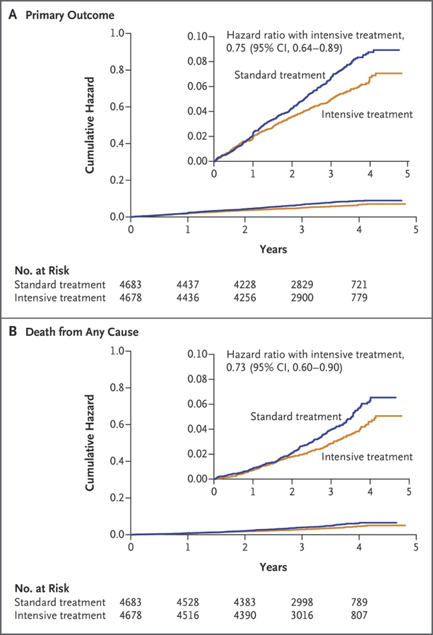
Treatment algorithm used all major classes of antihypertensives in no particular rank order. Participants were seen monthly for the first three months and every three months thereafter.  The median follow-up was 3.26 years, but the study was stopped early due to the benefit of treatment group by the data and safety monitoring board of the trial.


Outcomes

562 primary outcome events identified

  • 243 in the intensive group (1.65% per year)
  • 319 in the control group (2.19% per year)

Number needed to treat to prevent a primary outcome – 61

Number needed to treat to prevent death from any cause – 90

Number needed to treat to prevent death from cardiovascular causes – 172


Serious Adverse Events

1793 participants in intensive group (38.3%)

1736 participants in control group (37.1%)

Examples:

  • Hypotension
  • Syncope
  • Electrolyte abnormalities
  • Acute kidney injury
  • Injurious falls
  • Bradycardia


Discussion

25% lower relative risk of primary outcome in intensive group (5.2% vs 6.8%):

  • 38% lower relative risk of heart failure (1.3% vs 2.1%)
  • 43% lower relative risk of death from cardiovascular causes (0.8% vs 1.4%)
  • 27% lower relative risk of death from any cause (3.3% vs 4.5%)


SPRINT1

SPRINT Research Group. NEJM. 2015;373:2103-2116


SPRINT2

SPRINT Research Group.  NEJM. 2015;373:2103-2116

Should we be more aggressive in the SPRINT patient population?

(great review by Health News Review here)

First of all, this is only ONE study addressing a clinical question that has been extensively researched and we still don’t have good, reliable, reproducible results yet. See 2009 Cochrane Hypertension Review that stated blood pressure < 140/90 mmHg was not beneficial.

Second of all, the researchers seemed to down play the serious adverse events.  The author of the Health News Review put a nice table together and did a few extra EBM calculations (below) to help illustrate:

SPRINT-Table

125: Number needed to treat to prevent one case of heart failure.

167: Number needed to treat to prevent one death by cardiovascular causes

83: Number needed to treat to prevent death by any cause

100: Number needed to harm to cause one case of hypotension

167: Number needed to harm to cause one case of syncope

125: Number needed to harm to cause one case of electrolyte abnormality

56: Number needed to harm to cause one case of acute kidney injury or renal failure

42: Number needed to harm to cause one serious adverse event


So what I take away from the SPRINT study:

  1. This is data from a single study. Need more to change practice.
  2. Composite endpoints should be used with caution…especially since it was stopped early and may overestimate the benefit and underestimate the risk.
  3. Researchers used automated BP measurements which is shown to be lower (5-10 mmHg) than auscultated BP measurement. This alone could cause clinicians to intensify BP control and increase adverse events that weren’t needed.
  4. It doesn’t help us with patients with diabetes, previous stroke, symptomatic heart failure, or nursing home residents…which encompass a lot of patients being managed for hypertension.

References

  1. James PA, Oparil S, Carter BL, et al. 2014 evidence-based guideline for the management of high blood pressure in adults: report from the panel members appointed to the Eighth Joint National Committee (JNC 8). JAMA. 2014;311(5):507-20.
  2. Hernandez-Vila E. A review of the JNC 8 blood pressure guideline.  Tex Heart Inst J.  2015;42(3):226-228.
  3. The SPRINT Research Group. A randomized trial of intensive versus standard blood pressure control. N Engl J Med. 2015;373:2103-2116.

Fear and Education

This was my first blog post for The American Academy of Physician Assistants PA Blog in a recurring series called “Professor’s Corner”. 


           Fear is good.  It is a strong statement and is bound to invoke certain emotional feelings, which may not always be positive in nature.  Think back to the last time you were truly scared.  Chances are you can remember more than you think about the event in question.  Cognitive psychologists have studied the effect of fear on attention, as well as retention, and shows there may be a “sweet spot” for a “healthy amount” of stress that is beneficial to learners.  The trick is not only finding that perfect zone for each student, but also adjusting it throughout the 2.5 years of school. 

            The fear and stress at the beginning of the didactic phase of PA education is most commonly caused by grades and doing well on exams.  Many of you may have gone through undergraduate studies with very high marks, and maybe even didn’t have to study all that hard.  Then PA school hits you like a freight train.  So what happens?  You commit “academic bulimia”……binging on large amount of information for exams and then completely purging it from memory to make space for the next exam.  Very little retention takes place, but the fear of doing well in school is tempered.  Compare this to the end of the didactic year when you have your studying methods down, but you are preparing for clinical rotations and suddenly you think you “can’t remember anything”.  Now the fear is shifted from grades, to trying to remember as much as possible to take care of actual patients.  While on clinical rotations, your fear is redirected once again to trying to recall any information from your didactic year so you don’t look incompetent in front of your patients or preceptors.

            Our jobs as professors in PA programs is to instilling aliquots of fear in safe environments to get you ready to practice medicine.  Maybe it is using more simulation in a group setting.  Maybe it is calling on you in class.  Maybe it is pop quizzes.  Maybe it is comprehensive final exams.  The method to our madness……stress inoculation.  By doing small stressful tasks throughout your classroom instruction, we are trying to prepare you for learning in the clinical environment.  Simulation prepares you to make clinical decisions in low-stakes environments.  Calling on you in class prepares you for the Socratic method of teaching that permeates the halls of the hospitals.  Pop quizzes and comprehensive finals teaches you to be self-directed, intrinsic learners that will persist for your entire medical career.

              Fear is good.  No other profession has the life or death struggle that medicine embraces.  The moment you do not fear taking care of patients is the moment you may cause someone harm.  Part of the educational process of PA school is to teach you how to manage this fear.  Every time you overcome fear, you become stronger.  Suddenly, thinking about getting called out in class by a professor in a class of your peers is nothing compared to getting called on by your attending in front a group of strangers….or telling a patient they have terminal cancer.  Harnessing this fear and helping you focus it will not only help you be the best clinician possible, but it will indirectly help every one of your patients you take care for the rest of your career.

References

1)  Vermeulen N, Godefroid J, Mermillod M (2009) Emotional Modulation of Attention: Fear Increases but Disgust Reduces the Attentional Blink. PLoS ONE 4(11): e7924.

2)  Schwabe L, Joels M, Roozendaal B, Wolf OT, Oitzl MS.  Stress effects on memory:  An update and integration.  Neuroscience and Biobehavioral Reviews.  2012;36:1740-1749.

3)  Susskind JM, Lee DH, Cusi A, Feiman, Grabski W, Anderson AK.  Expressing fear enhances sensory acquisition.  Nature Neuroscience.  2008;11(7):843-850.

4)  Perry B.  Fear and Learning: Trauma-Related Factors in Adult Education Process.  New Directions for Adult and Continuing Education.  2006;110:21-27.

#5 – Aortic Dissections


***Listen to podcast by clicking here***


Cliff Reid - SMACC 2015

Cliff Reid – SMACC 2015

Background

  • 1st described by German anatomist Daniel Sennert in 16th century on autopsy
  • King George II of England died of aortic dissection in 1760 and described by Frank Nichols
  • John Ritter (Actor) died of dissection
  • Relatively uncommon, but can be fatal if missed
  • 3-5 cases/100,000 each year
  • Mortality rates around 25%
  • 22% undiagnosed prior to death

Pathophysiology

  • Tear in aortic intima that leads to false lumen between intima and media
    • majority occur in ascending aorta between the sinotubular junction and left subclavian artery
  • Bimodal age distribution
    • Teens-30yo and > 50yo

Risk Factors

  • Chronic hypertension
  • Connective tissue disorders
    • Marfans, Ehler-Danlos
  • Bicuspid aortic valve
  • Previous aortic instrumentation
  • Family history of dissections

Classification

  • DeBakey (older)
    • Uses site of origin
      • Type I
        • Originates in ascending and includes the arch
      • Type II
        • Originates and confined to ascending aorta
      • Type III
        • Originates in descending and extends proximal/distal
  • Stanford (more widely used)
    • Stanford A
      • Involves the ascending aorta
    • Stanford B
      • Everything else
  • Newer DISSECT classification can be used to help with treatment options
    • Duration
      • Acute = < 2 weeks
      • Subacute = 2 weeks to 3 months
      • Chronic = > 3 months
    • Intimal Tear
      • Primary location
        • Ascending
        • Aortic Arch
        • Descending
        • Abdomen
    • Size of Aorta
      • Maximum trans-aortic diameter within dissected segment
    • Segmental Extent of aortic involvement
      • Broken down in to sections from arch to iliacs
    • Clinical complications associated with dissection
    • Thrombus of aortic false lumen
      • Patent or not

Clinical Presentations

  • History
    • Pain (90%)  – sharp > ripping/tearing
      • Chest in Type A (83%)
      • Back (64%) and abdominal (43%) in Type B
    • Neurologic
      • Syncope, stroke symptoms (more common in Type A)
      • Type B – paraplegia from Artery of Adamkiewicz
      • Horner’s Syndrome from compression on superior cervical ganglion
      • Hoarseness from compression on left recurrent laryngeal nerve
  • Physical Exam
    • Tamponade (most common cause of death) in Type A or Type I
      • Beck’s Triad
        • Hypotension, JVD, muffled heart tones
    • Can be hypertensive or hypotensive
    • Pulse deficits (> 20mmHg variation)
      • Higher mortality if present
      • Depends on the site (up to 30% of Type A vs 10% of Type B)
      • Older patients less likely to have pulse discrepancy
    • Heart murmur (more common in younger patients)
      • Aortic regurgitation (50-66%)
        • Diastolic decrescendo murmur
        • Heard best over right sternal border
          • as opposed to classic primary aortic disease AR which is heard over the left sternal border
  • Pre-test Probability
    • 77% of patients have 2 out of the 3 high-risk variables:
      • Variation in pulse or blood pressure
      • Presence of CXR abnormality
      • Abrupt onset of sharp, chest/abdominal pain

Diagnostic Studies

  • EKG (almost always 1st study done for chest pain)
    • Not very helpful other than to R/O ACS
      • May be normal in 19-31% of dissections
    • May see abnormal changes if dissection involves coronary arteries
  • Chest Radiograph
    • Classic finding is widening of the mediastinum or aortic silhouette
      • Incidence ~ 60%
    • Up to 30% of patients with aortic dissection have no CXR abnormality

  • D-Dimer
    • Extensively studied…but essentially worthless
      • When < 500 ng/mL:
        • Sensitivity – 97%, Negative Predicative Value – 96%
        • Specificity – 56%
        • False negative rate of 18%
  • Computed Tomography Angiography (hemodynamically stable)
    • Test of choice for diagnosis
    • Findings of acute dissection:
      • Intimal flap
      • True and false lumen
      • Pericardial effusion
  • Echocardiography (hemodynamically unstable)
    • Transesophageal (preferred)
      • Sensitivity/specificity can approach CT numbers
      • Require procedural sedation
    • Transthoracic (acceptable)
      • Quicker, no sedation, inferior sensitivity/specificity to CT

Intimal Flap

Treatment

  • Acute Management
    • Control of heart rate and blood pressure
      • Systolic BP < 120 mmHg
      • Heart rate < 60 bpm
    • Medications
      • IV Beta-blocker (1st line)
        • Esmolol (0.1-0.5 mg/kg over 1 minute followed by 0.025-0.2 mg/kg/hr)
        • Labetalol (20mg bolus followed by 0.5-2 mg/hr)
        • Propanolol (1-10mg load followed by 3 mg/hr)
      • If hypertensive after beta-blockade:
        • Nitroprusside (0.25-0.5 mcg/kg/min)
        • Nicardipine (5mg/hr, increase 2.5 mg/hr q15min to max 15mg/hr)
  • Definitive Management
    • Type A (surgical emergency)
      • Mortality rate 1-2% per hour after symptom onset
      • Open repair on bypass
        • Median sternotomy with graft placement +/- aortic valve replacement
        • May need to re-implant coronary and/or great vessels
      • > 90% 3yr survival after surgery


  • Type B
    • Uncomplicated
      • Medical therapy
        • BP goal = < 120/80 mmHg
        • Oral beta-blocker is 1st line
        • Add ACE inhibitors or calcium channel blocker to BP goal
      • Serial Imaging
        • CT or MRI at 3, 6, and 12 months
        • If no progression, then every 1-2 years
      • Complicated (or progressing)
        • Endovascular graft surgery
          • Indications
            • Involvement of major aortic branch leading to end organ ischemia
            • Persistent HTN or pain
            • Aneurysmal dilation
            • Concomitant connective tissue disorder


References

  1. Sennertus D: Cap. 42, Op Omn Lib. 5:306-315, 1650
  2. Nichols F. Observations concerning the body of his late majesty, October 26, 1760, Phil Trans Lond. 52:265-274, 1761.
  3. Pacini D, Di marco L, Fortuna D, et al. Acute aortic dissection: epidemiology and outcomes. Int J Cardiol. 2013;167(6):2806-12.
  4. Hagan PG, Nienaber CA, Isselbacher EM, et al. The International Registry of Acute Aortic Dissection (IRAD): new insights into an old disease. JAMA. 2000;283(7):897-903.
  5. Olsson  C, Thelin  S, Stahle  E  et al.: Thoracic aortic aneurysm and dissection: increasing prevalence and improved outcomes reported in a nationwide population-based study of more than 14,000 cases from 1987-2002. Circulation 114: 2611, 2006.
  6. E. DeBakey, W.S. Henley, D.A. Cooley, G.C. Morris, E.S. Crawford, A.C. Beall. Surgical management of dissecting aneurysms of the aorta.  J Thorac Cardiovasc Surg. 1965;49:130–149
  7. O. Daily, H.W. Trueblood, E.B. Stinson, R.D. Wuerflein, N.E. Shumway. Management of acute aortic dissections.  Ann Thorac Surg. 1970;10:237–247.
  8. Dake MD, Thompson M, Van sambeek M, Vermassen F, Morales JP. DISSECT: a new mnemonic-based approach to the categorization of aortic dissection. Eur J Vasc Endovasc Surg. 2013;46(2):175-90.
  9. Dake MD, Thompson M, Van sambeek M, Vermassen F, Morales JP. DISSECT: a new mnemonic-based approach to the categorization of aortic dissection. Eur J Vasc Endovasc Surg. 2013;46(2):175-90.
  10. Pape, L. A., Awais, M., Woznicki, E. M., Suzuki, T., Trimarchi, S., Evangelista, A., Myrmel, T., Larsen, M., Harris, K. M., Greason, K., Di Eusanio, M., Bossone, E., Montgomery, D. G., Eagle, K. A., Nienaber, C. A., Isselbacher, E. M., & O’Gara, P. Presentation, Diagnosis, and Outcomes of Acute Aortic Dissection: 17-Year Trends From the International Registry of Acute Aortic Dissection. Journal of the American College of Cardiology. 2015;4:350–358.
  11. Mehta, R. H., O’Gara, P. T., Bossone, E., Nienaber, C. A., Myrmel, T., Cooper, J. V., Smith, D. E., Armstrong, W. F., Isselbacher, E. M., Pape, L. A., Eagle, K. A., Gilon, D. Acute type A aortic dissection in the elderly: clinical characteristics, management, and outcomes in the current era. Journal of the American College of Cardiology. 2002;4:685–692.
  12. Movsowitz, H. D., Levine, R. A., Hilgenberg, A. D., & Isselbacher, E. M. Transesophageal echocardiographic description of the mechanisms of aortic regurgitation in acute type A aortic dissection: implications for aortic valve repair. Journal of the American College of Cardiology. 2000;3:884–890.
  13. Biagini E, Lofiego C, Ferlito M, et al. Frequency, determinants, and clinical relevance of acute coronary syndrome-like electrocardiographic findings in patients with acute aortic syndrome. Am J Cardiol. 2007;100(6):1013-9
  14. Von kodolitsch Y, Schwartz AG, Nienaber CA. Clinical prediction of acute aortic dissection. Arch Intern Med. 2000;160(19):2977-82.
  15. Hogg K, Teece S. Best evidence topic report. The sensitivity of a normal chest radiograph in ruling out aortic dissection. Emerg Med J. 2004;21(2):199-200.
  16. Shimony A, Filion KB, Mottillo S, Dourian T, Eisenberg MJ. Meta-analysis of usefulness of d-dimer to diagnose acute aortic dissection. Am J Cardiol. 2011;107(8):1227-34.
  17. Sueyoshi E, Nagayama H, Hayashida T, Sakamoto I, Uetani M. Comparison of outcome in aortic dissection with single false lumen versus multiple false lumens: CT assessment. Radiology. 2013;267(2):368-75.
  18. Moore AG, Eagle KA, Bruckman D, et al. Choice of computed tomography, transesophageal echocardiography, magnetic resonance imaging, and aortography in acute aortic dissection: International Registry of Acute Aortic Dissection (IRAD). Am J Cardiol. 2002;89(10):1235-8.
  19. Hiratzka LF, Bakris GL, Beckman JA, et al. 2010 ACCF/AHA/AATS/ACR/ASA/SCA/SCAI/SIR/STS/SVM guidelines for the diagnosis and management of patients with Thoracic Aortic Disease: a report of the American College of Cardiology Foundation/American Heart Association Task Force on Practice Guidelines, American Association for Thoracic Surgery, American College of Radiology, American Stroke Association, Society of Cardiovascular Anesthesiologists, Society for Cardiovascular Angiography and Interventions, Society of Interventional Radiology, Society of Thoracic Surgeons, and Society for Vascular Medicine. Circulation. 2010;121(13):e266-369.
  20. Tsai TT, Nienaber CA, Eagle KA. Acute aortic syndromes. Circulation. 2005;112(24):3802-13.
  21. Trimarchi S, Nienaber CA, Rampoldi V, et al. Contemporary results of surgery in acute type A aortic dissection: The International Registry of Acute Aortic Dissection experience. J Thorac Cardiovasc Surg. 2005;129(1):112-22.