Answer to Heme/Onc Questions

Question #1

Which of the following genetic mutations is seen with chronic myelogenous leukemia?

Answer:

Translocation of chromosome 9 and 22.  This new chromosome 22 is called “Philadelphia chromosome” after the city where the two hospitals that first identified the gene mutation in 1960 were both located.

Question #2

Which of the following cancer markers are classically associated with ovarian cancer?

Answer:

CA-125

cancertumormarkersdiagram

#10 – Approach to Anemia in Adults


Definition

The textbook definition of anemia is a reduction of the absolute number or mass of circulating red blood cells.  This then causes a global reduction in the oxygen carrying capacity of the patient’s circulatory system.  Clinically, we use hemoglobin and hematocrit as the surrogate markers and define anemia as 2 SD below the mean for gender:

  • Men
    • Hemoglobin < 13.5 g/dL
    • Hematocrit < 41%
  • Women
    • Hemoglobin < 12.0 g/dL
    • Hematocrit < 36%


tileshop.fcgi

Patel KV. Haematologica. 2008;93(9):1281-1283.


  • Special Populations
    • Athletes
      • May have a baseline anemia due to:
        • Dilution from increased plasma volume
        • Hemolytic breakdown from exercise
        • Exercise induced cytokines decreases RBC production
      • A normal H/H in a competitive athlete may suggest performance enhancing drugs
    • High altitudes
      • May have elevated hemoglobin concentration as baseline
    • Smokers
      • Baseline higher hemoglobin due to carboxyhemoglobin

General Causes of Anemia

There are two general approaches you can use to help identify the cause of anemia in adults.

  • The Kinetic Approach (the mechanisms responsible for the low hemoglobin)
    • 3 independent mechanisms
      • Decreased RBC production
        • Lack of nutrients
        • Bone marrow failure
        • Decreased erythopoetic stimulation factors
          • Erythropoietin, T3, androgens
        • Inflammation
      • Increased RBC destruction
        • Hemolysis, hypersplenism
      • Blood loss
  • The Morphologic Approach (categories based on RBC size and reticulocyte response)
    • Macrocytic (MCV > 100 fL)
      • Vitamin B12, folate, EtOH, liver disease
      • Any condition causing reticulocytosis
    • Microcytic (MCV < 80 fL)
      • 3 most common in clinical practice
        • Iron deficiency
          • ↓ serum iron, ↓ serum ferritin, ↑ TIBC
        • Alpha or beta thalassemia minor
          • Normal iron studies
        • Anemia of chronic disease
          • ↓ serum iron, normal serum ferritin, ↓ TIBC

History Questions

  • Is the patient symptomatic?
    • Fatigue, dyspnea, bleeding, bruising, dizziness, syncope
  • Any history of weight loss, night sweats, fever, anorexia?
    • Infection or malignancy
  • Past medical history for chronic illness
    • PUD, renal disease, autoimmune conditions, liver disease, past malignancies
  • Family history for hemoglobinopathies
  • Social history for alcohol use
  • Occupational exposures

Physical Exam Findings

  • Pallor
    • Palms, nail beds, face, conjunctiva
  • Jaundice
  • Hepatosplenomegaly
  • Lymphadenopathy
  • Petechiae, purpura, bruising
  • Bone tenderness

Laboratory Testing

Anemia is usually first diagnosed by CBC.  Once you have a documented low H/H, then you need order follow-up studies to help differentiate the cause of the anemia.  These include:

  • RBC indices
    • MCV, MCH, MCHC, RDW
  • Reticulocyte count and index
  • Peripheral smear
    • Helmet cells or schistocytes à microangiopathic hemolysis
    • Microspherocytes à autoimmune hemolysis
    • Tear drop RBC à myelofibrosis
    • Bite cells à oxidative hemolysis
    • Parasites à malaria, babeosis
    • Hypersegmented neutrophils à Vitamin B12 or folate deficiency
    • Nucleated RBC
    • Siderocytes
    • Target cells à thalassemias
  • WBC and platelet count from CBC
  • If hemolysis is suspected:
    • ↑ Serum LDH, ↓ serum haptoglobin, and ↑ serum indirect bilirubin
    • Direct Coombs test (antibodies against RBC)
  • Bone marrow evaluation
Picture1

Schrier SL, et al. Approach to adults with anemia. In: Up To Date. Waltham, MA (Accessed 03/23/2016)

The Cottage Physician Management

Something new I thought I would bring to the PAINE Podcast.  As you all know, I am quite a fan of medicine and antiquity.  Shortly after I married my wife, her grandfather past away from a progressive esophageal cancer.  One of the things I was able to keep when helping clean out his house, was a copy of The Cottage Physician printed in 1893.  It was basically a handbook on how to treat common ailments of the time. I will try to add excerpts from this book when appropriate so you can have a sense of how medicine was practiced in the late 19th century.

Cottage Physician - Anemia

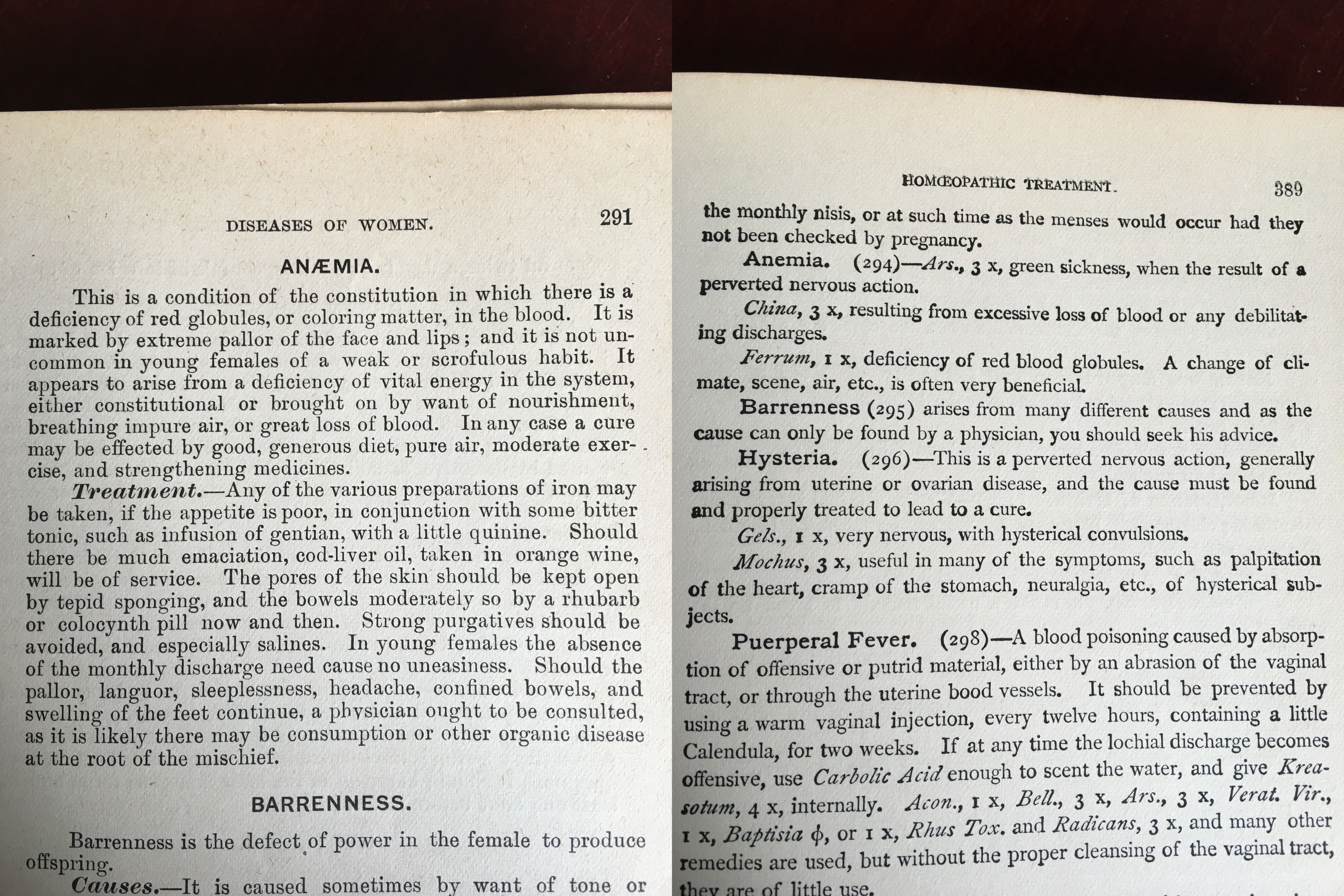
The Cottage Physician. 1863

References

  1. Patel KV. Variability and heritability of hemoglobin concentration: An opportunity to improve understanding of anemia in older adults.  Haematologica.  2008;93(9):1281-1283.
  2. Beutler E, Waalen J. The definition of anemia: what is the lower limit of normal of the blood hemoglobin concentration?. Blood. 2006;107(5):1747-50.
  3. Shaskey DJ, Green GA. Sports haematology. Sports Med. 2000;29(1):27-38.
  4. Ruíz-argüelles GJ. Altitude above sea level as a variable for definition of anemia. Blood. 2006;108(6):2131.
  5. Nordenberg D, Yip R, Binkin NJ. The effect of cigarette smoking on hemoglobin levels and anemia screening. JAMA. 1990;264(12):1556-9.
  6. Hillman RS, Ault KA, Leporrier M, Rinder HM. Clinical Approach to Anemia.  In: Hematology in Clinical Practice.  5th McGraw-Hill. New York. 2010.
  7. Tefferi A. Anemia in adults: a contemporary approach to diagnosis. Mayo Clin Proc. 2003;78(10):1274-80.
  8. Nardone DA, Roth KM, Mazur DJ, Mcafee JH. Usefulness of physical examination in detecting the presence or absence of anemia. Arch Intern Med. 1990;150(1):201-4.
  9. Hung OL, Kwon NS, Cole AE, et al. Evaluation of the physician’s ability to recognize the presence or absence of anemia, fever, and jaundice. Acad Emerg Med. 2000;7(2):146-56.

Answer to Hematology/Oncology Case #1

Answer:

Check a methylmalonic acid level


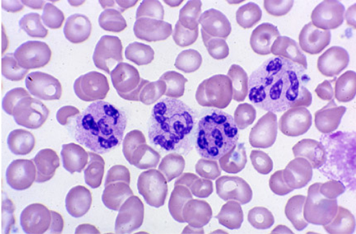
This patient has had a history of gastric cancer with a partial gastrectomy and now presents with a fatigue and gait disturbances.  CBC reveals a macrocytic aneamia and peripheral smear shows multinucleated neutrophils.  The gait disturbances are most likely due to the progressive peripheral neuropathy.  This is most consistent with vitamin B12 deficiency.  Intrinsic factor, which is secreted by the parietal cells of the stomach, is required for vitamin B12 absorption in the terminal ileum.

Work-Up for Vitamin B12 Deficiency

Serum B12 Level

  • < 300 pg/dL is diagnostic

Metobolites

  • Methylmalonic acid
    • < 70 nanomol/L is diagnostic
  • Homocysteine
    • < 5 micromol/L is diagnostic

Possible additional testing in the setting of macrocytic anemia:

  • If pernicious anemia is suspected:
    • Anti-intrinsic factor antibodies
  • If folate deficiency is suspected:
    • Serum folate level
      • < 2 ng/mL is diagnostic
    • RBC folate level (reserved for indeterminate serum levels)
      • < 280 ng/mL is diagnostic

Treatment for B12 Deficiency

  • Intramuscular
    • 1mg daily x 7 days, then 1mg weekly for 4 weeks, then 1mg monthly
  • Oral
    • 1000-2000mg daily

References

  1. Antony AC. Megaloblastic anemias. In: Hematology: Basic principles and practice, 4th ed, Hoffman R, Benz EJ, Shattil SJ, et al. (Eds), Churchill Livingstone, New York 2005. p.519.
  2. Butler CC, Vidal-alaball J, Cannings-john R, et al. Oral vitamin B12 versus intramuscular vitamin B12 for vitamin B12 deficiency: a systematic review of randomized controlled trials. Fam Pract. 2006;23(3):279-85.

Hematology/Oncology Case #1

62-year-old male presents to primary provider’s office with a six-month history of fatigue and gait disturbance.  He denies recent falls, weakness, pain, paralysis, or dizziness.


Medications

Lisinopril 10mg daily

Metformin 1000mg BID

Men’s multivitamin

Fish oil


Past Medical History

Diabetes Mellitus II

Hypertension

Gastric cancer


Past Surgical History

Cholecystectomy – 1997

Partial gastrectomy – 2004


Vitals

BP-128/79, HR-81, RR-14, O2-100%, Temp-98.9o


Physical Exam

General – WN/WD, NAD

Skin – scattered senile purpura, no petechiae

CV – RRR without M/G/R

Pulmonary – CTA bilaterally without adventitial breath sounds

Neurologic – A&Ox3, 5/5 strength throughout bilaterally, DTR 2+ and equal, FROM, vibratory sensation decreased in bilateral lower extremities


Laboratory Studies

Case#1

Hypersegmented Neutrophils


#9 – The CBC Demystified


***Listen to the podcast here***


White Blood Cell Count

  • Normal range (4.0-11.0 x 10³ cells/mm3)
  • 2 Parts
    • Actual count of the number of leukocytes in one cubic millimeter of blood
    • Differential count (% of each of the 5 types of leukocytes)
  • < 4.0 x 10³ cells/mm3 = leukopenia
    • Bone marrow failure, blood malignancies, infections
  • > 11.0 x 10³ cells/mm3 = leukocytosis
    • Infections, inflammation, blood malignancies
  • Differential
    • Granulocytes
      • Neutrophils (45-73%)
        • Bands (3-5%)
        • “Left shift”
        • Absolute Neutrophil Count (ANC)
          • (%PMN + %Bands) x WBC / 100
          • < 1500 = neutropenia
      • Eosinophils (0-4%)
      • Basophils (0-1%)
    • Non-granulocytes
      • Lymphocytes (20-40%)
        • 3 Main Types
          • T-Cells = cell mediated immunity
          • B-Cells = humoral immunity (immunoglobulins)
          • Natural Killer Cells (NKC)
      • Monocytes (2-8%)

Never Let Monkeys Eat Bananas

Red Blood Cell Count

  • Actual count of RBC in one cubic millimeter of blood
    • Normal range
      • Male (4.5-5.9 x 10¹² cells/L)
      • Female (4.1-5.1 x 10¹² cells/L)
    • Reasons for gender differences in RBC markers
      • Blood loss from menstruation
      • Testosterone is hematapoetic
    • 2 main markers
      • Hemoglobin
        • Amount of Hgb contained in a given volume of blood
        • Normal Range
          • Male (14-17.5 g/dL)
          • Female (12.3-15.3 g/dL)
      • Hematocrit
        • % of total blood volume made up by RBC
        • Normal range
          • Male (42-50%)
          • Female (36-45%)
        • As a general rule of thumb, most agreed upon transfusion trigger is Hgb < 7.0 g/dL and/or Hct < 20%

Red Blood Indices

  • Essentially assess the size and hemoglobin content of the RBC and is useful in the evaluation of anemias, polycythemias, and nutritional deficiencies
  • Mean Corpuscular Volume (MCV)
    • Average size of the volume of RBC (Hct/RBC count)
    • Normal range (80-100 fL/cell)
      • < 80 = Microcytic
      • > 100 = Macrocytic
  • Mean Corpuscular Hemoglobin (MCH)
    • Average amount of hemoglobin within a RBC
    • Closely resembles MCV and of little clinical use
  • Mean Corpuscular Hemoglobin Concentration (MCHC)
    • Calculation of Hgb/Hct
    • Normal range (32-36 g/dL)
      • < 32 = hypochromic
      • > 36 = hyperchromic
  • Red Blood Cell Distribution Width (RDW)
    • Indication of RBC size variability (degree of anisocytosis)
    • Normal range (11-14%)
  • Reticulocyte Count
    • Measure of immature RBC in circulation
      • Evaluates bone marrow function and erythropoetic activity
    • Normal range (0.5-2.5%)
    • Reticulocyte Index
      • Measures whether this response is appropriate
      • Reticulocyte Count % x (Patient’s Hct/Normal Hct)
        • < 1.0 = poor response

Platelet Count

  • Actual count of thrombocytes in one cubic millimeter of blood
  • Normal range (150,000-450,000/µL)
  • Mean Platelet Volume (MPV)
    • Relationship between size and count
    • Immature platelets are larger
    • MPV is analogous to the MCV

Strategies for Quick Interpretation

  • If abnormal WBC:
    • Correlate with clinical picture
      • An abnormal number may be a normal response to illness or a marker of illness severity
    • Look at your differential
    • If leukopenic, then calculate ANC
  • If abnormal RBC:
    • Is your patient symptomatic?
    • Look at your indices
    • Calculate reticulocyte count for response
  • If abnormal platelet:
    • Is the patient bleeding?
Untitled-1

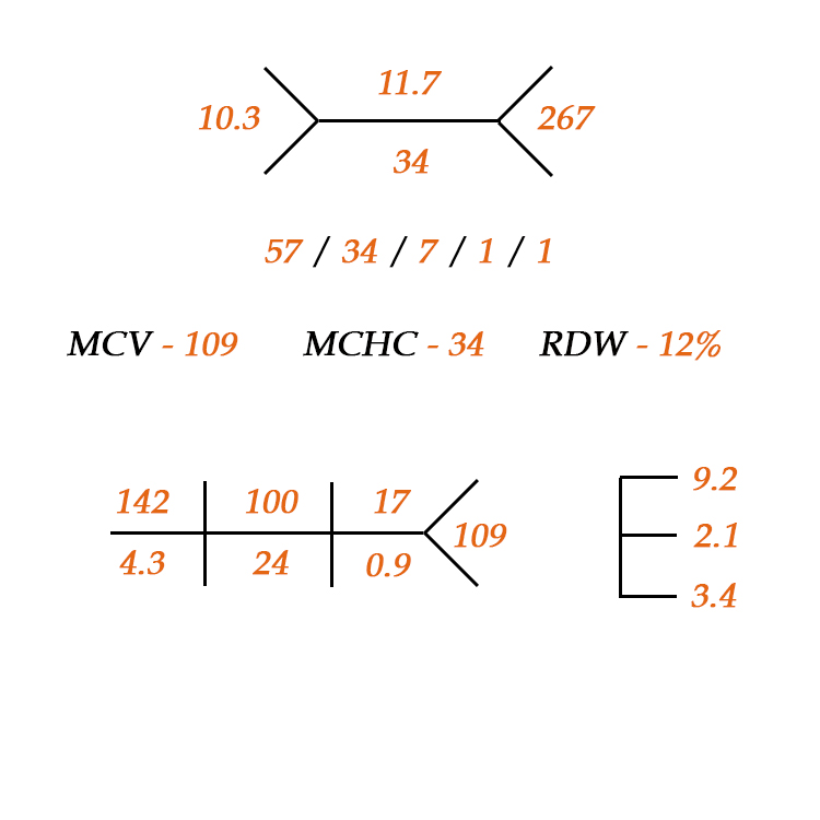
Fishbone diagram for CBC with differential

References

1) Pagana KD, Pagana TJ.  Mosby’s Manual of Diagnostic and Laboratory Tests.  5th ed.  St. Louis, MO. Elsevier. 2014.

2) Lee M.  Basic Skills in Interpreting Laboratory Data.  5th ed.  Bethesda, MD.  American Society of Health-Systems Pharmacists.  2013

3) Laposata M.  Laboratory Medicine: The Diagnosis of Disease in the Clinical Laboratory.  2nd ed.  New York, NY.  2014.

Memory and Learning

This is my post for The American Academy of Physician Assistants Blog “PAs Connect” in a recurring series called “Professor’s Corner”.


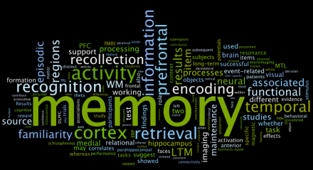
School is all about memory.  You are presented information in class and you try to remember everything the professor is saying in order to transcribe what you think is important on your notes.  Then you go home and study these notes, textbook chapters, and journal articles to help try to make sense of the material. This is where our cognitive weapons of choice come out: different colored highlighters, index cards, sticky notes….all in the hopes that it is processed into neat little compartments in your brain so you can recall them for the exam.  Rinse…wash…repeat for 2.5 years of PA school.  But what is the science behind memory and why do somethings work better than others?

Most people think of memory as just short term and long term, but depending on which psychologic theory of memory you prescribe to, there can be many more.  I, personally and professionally, like the three-tiered, information processing model to memory: sensory, working, and long-term.

Memory Graphic

Sensory Memory

The first step in memory involves the processing of sensory stimuli that are introduced in the learning environment.  The brain uses three main senses (sight, sound, touch) to get as much information brought in as possible.  Now, if you had to process every single sensory stimulus into your working memory, your brain would get overloaded in a matter of seconds.  To avoid this, you use sensory memory as the environmental buffer to pick and choose what you think is the most important……this is where attention comes in.  Attention is the filter from the sensory memory system into the working memory system and allows us to focus on smaller, more important sensory stimuli.

Attention is the bodyguard outside of Club Working Memory.  He will pick and choose what information gets through for further processing to make sure the club isn’t overcrowded.

Working Memory

Once the sensory information is attended to, we can store it in a temporary cognitive sandbox where we can actually work with it.  This is your working memory.  It is short (15-30s) and the information stored here only stays if the learner is actually doing something with it (rehearsal).  There are 3 main components of working memory:  central executive (supervisor), the phonological loop (language storage), the visuospatial sketchpad (visual storage).  As you hear and see things over and over again, you start to make connections and correlation between the information and begin to encode it all into neat data packages.  Each system is different and the cognitive load when using both systems is only slightly higher than using them individually.  Cognitive psychologists recommend using this to your advantage while studying.  For example, assign each class a room in your house and only study in this room.  It will increase the likelihood of correctly recalling the information when you can get your cognitive bearings straight on the location you learned the information.  You can incorporate similar strategies such as listening to different music or artists for different subjects.

The more you rehearse this information in working memory, the greater the cognitive load….but the higher likelihood it will make it to long-term memory.

Working Memory Graphic

Long-term Memory

Due to this increased cognitive load of working memory, it is not possible to keep information in working memory indefinitely.  This is where long-term memory comes into play.  We try to categorize this information into packages for easy storage and recall in long-term memory.  There are 3 steps to this process:  encoding, storage, and retrieval/rehearsal.  The majority of the energy used when studying is trying to encode the information in working memory so it can be efficiently and effortless recalled when needed.  This is why mnemonics, acronyms, and all the other things we do as students help process and package new information so we can remember them later.  “Oh Oh Oh To Touch And Feel Very Good Velvet, Ahhhh” may mean nothing to you, but it is how I can still recall all 12 cranial nerves to this day.

Loss of Memory

Even with all these sophisticated and complicated cognitive pathways that our brains have developed to retain information, we still forget and lose information.  There are many theories behind this, but the 2 main ones I like are:  information decay (the less you use it, the less you can recall) and interference (learning new information inhibits recall of old information).

To limit interference when I am teaching, I try not to overload the senses with extraneous stimuli.  If I stand up and just read the powerpoint (which is not teaching by the way), you (the student) can’t listen to what I am saying when you are being bombarded with paragraphs of text that you feel compelled to read. This leads to overload of the sensory and working memory as you are trying to listen, read, and process everything that is being thrown at you.

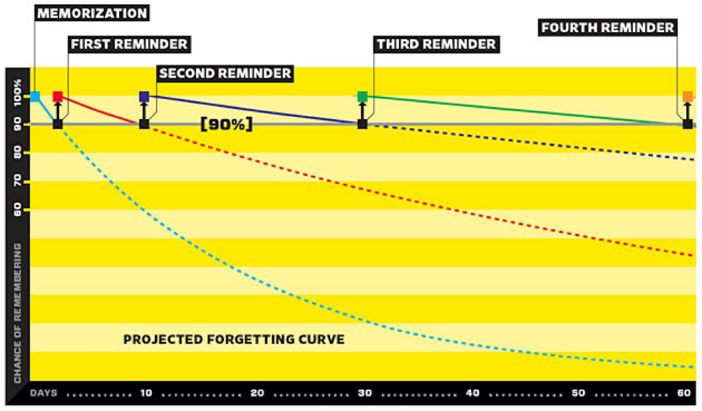
To limit long-term memory decay, I utilize spaced repetition.  When I am teaching, I like to bring up old information (last exam, least semester, etc…) to re-introduce to it to my students.  It is also why I believe in comprehensive final examinations.  It gives them an opportunity to dust off the stored information in long-term memory and play with it again in the sandbox of working memory.  This strategy has been shown to improve retention and efficiency of recall when you need it the most…..when taking care of patients.

SPaced Repitition

Wolf G. Want to Remember Everything You’ll Ever Learn? http://www.wired.com/2008/04/ff-wozniak/?currentpage=all


References

1)  Anderson JR.  Cognitive psychology and its implications.  7th ed.  Worth Publishers. 2009

2) McLeod SA. Working Memory | Simply Psychology. Available at: http://www.simplypsychology.org/working memory.html. Accessed February 15, 2016.

3) Cognition (Sensory memory). Introduction to Instructional Design. Available at: http://byuipt.net/564/2013/08/23/cognition-sensory-memory/. Accessed February 15, 2016

4)  Baddeley AD.  Working memory.  Science.  1992;255:556-559.

5) Cognition (Long-term memory). Introduction to Instructional Design. Available at: http://byuipt.net/564/2013/08/23/cognition-long-term-memory/. Accessed February 15, 2016.

6) Wolf G. Want to Remember Everything You’ll Ever Learn? Surrender to This Algorithm. Wired.com. Available at: http://www.wired.com/2008/04/ff-wozniak/?currentpage=all. Accessed February 15, 2016.

7) Nickson C. Learning by Spaced Repetition. Life in the Fast Lane Medical Blog 2011. Available at: http://lifeinthefastlane.com/learning-by-spaced-repetition/. Accessed February 15, 2016

Answer to OB/GYN Case #2

Answers

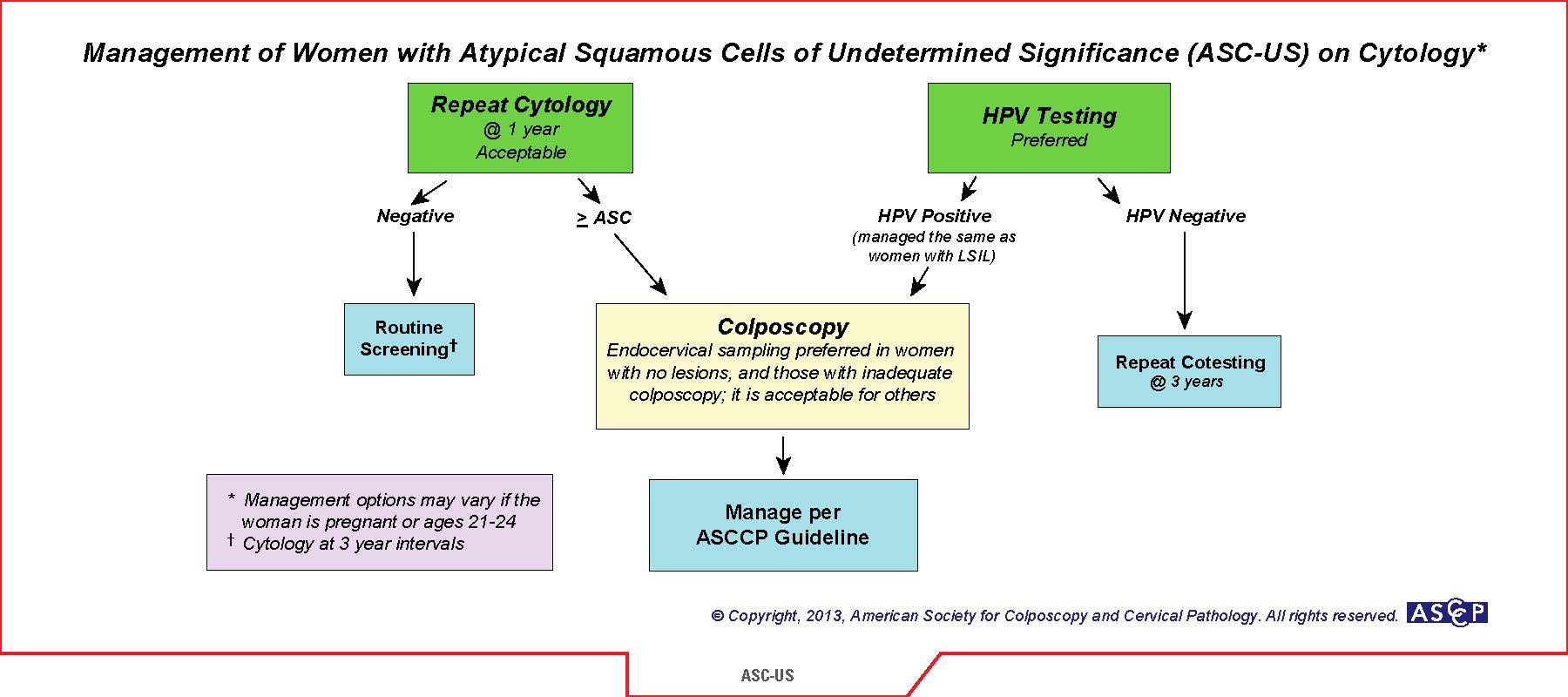
Next best step: Reflex HPV cotesting

Follow-up Recommendations: 3 years

Discussion

Atypical squamous cells of undetermined significance (ASC-US) is a common “abnormal” pap result.  The 2012 American Society of Clinical Pathologist (ASCP) recommend that women ages 21-29 should have routine cytology alone performed every 3 years as long as the results are normal.  If ASC-US results, then reflex HPV contesting is recommended.  If HPV (-), then return to routine cytology in 3 years.  If HPV (+), then proceed to colposcopy.  Another acceptable option is to have the patient return to clinic in 1 year for repeat pap.  If ASC-US (+) again, then proceed to colposcopy.  The logic is that most women in this age group clear any HPV infection without the need for colposcopy.  This is the safer choice if the patient is wanting to have more children to limit any complications of cervical incompetence.

ASCCP Management Guidelines_August 2014_Page_07

References

1)  American Society of Clinical Pathologists. Screening Guidelines. Available at: http://www.asccp.org/guidelines/screening-guidelines.


#8 – Gestational Hypertension


****Listen to the podcast by clicking here****


Definition of Gestation/Pregnancy-Induced Hypertension

  1. Any new onset (not previously diagnosed) of hypertension (SBP > 140mmHg and/or DBP > 90mmHg) at > 20 weeks gestation in the absence of proteinuria or new signs of end-organ damage
    1. Severe = SBP > 160mmHg and/or DBP > 110mmHg
  2. Documented on at least 2 occasions at least 4 hours apart

Epidemiology

  • 5-10% of all pregnancies
    • 6-17% of healthy nulliparous women
    • 2-4% of healthy multiparous women
  • 16% of all maternal deaths are related to hypertensive disorders

Pathophysiology

This is still unknown but several theories exist and include:

  • Maladaption to the normal physiologic changes of pregnancy
    • Increased blood volume
    • Elevated angiotensinogen from increased estrogen production
  • Abnormal trophoblast invasion of uterine blood vessels
    • Causes spiral arterioles to narrow
  • Immunologic intolerance between fetoplacental and maternal tissues

Risk Factors

  • Previous history of preeclampsia
  • Multifetal gestation
  • Overweight/obese
  • African-American

Fetal Well-being

  • Non-stress test and ultrasound should be performed upon diagnosis to assess fetal growth, fetal measurements, and amniotic fluid estimation and serially depending on severity.

Laboratory Evaluation

  • 24-hour urine collection for protein
    • > 300mg of protein = proteinuria
  • Platelet count
    • < 100,000 = thrombocytopenia
  • Liver Function Test
    • 2x transaminases = impaired liver function
  • Serum creatinine
    • > 1.1mg/dL = impaired renal function

Preeclampsia

  • Gestational hypertension with proteinuria and/or end-organ damage
    • Pulmonary edema, cerebral or visual disturbance, or any of the above laboratory abnormalities
  • 10-50% of women with gestation hypertension go on to develop preeclampsia within 5 weeks of diagnosis
  • Risk factors
    • Gestational hypertension diagnosed < 34 weeks gestation
    • Mean SBP > 135mmHg on 24-hour monitoring
    • Abnormal uterine artery Doppler
    • Elevated serum uric acid level (> 5.2mg/dL)

Management

Revolves around 3 main factors:

  1. Fetal growth and maturation
  2. Maternal and fetal benefits from early intervention
  3. Maternal and fetal risk from expectant management
HYPITAT

Broekhuijsen K, et al. Lancet. 2015;385(9986):2492-501.

HYPITAT-II Trial (HYPertension and Preeclampsia Intervention At Term)

  • 897 women diagnosed with non-severe gestational hypertension between 34-37 weeks gestation
  • Study group – delivery within 24-hours of diagnosis (induction or cesarean)
  • Control group – management until 37 weeks gestation
  • Results
    • 3% of control group vs 0% of study group developed at least one of the following:
      • Thromboembolic complications
      • HELLP syndrome
      • Eclampsia
      • Placental abruption
    • 3% of of study group developed neonatal respiratory distress syndrome vs 1.1% of control group

Non-severe (<160/110mmHg) and no preeclampsia

  • Screening
    • BP monitoring one or twice weekly with weekly assessment of proteinuria, platelet count, and liver enzymes
    • Weekly NST with sonographic estimation of amniotic fluid index
  • No evidence for starting antihypertensive therapy, unless patient has pre-existing end-organ dysfunction that could be worsened with hypertension (renal, cardiac, etc.)
  • Plan for delivery between 37-38 weeks

Severe (>160/110mmHg) and no preeclampsia

  • Same screening recommendations
  • Should be treated with antihypertensive therapy

Meds

  • Goals
    • No evidence of end-organ damage = < 160/110mmHg
    • Evidence of end-organ damage = < 140/90mmHg
  • Corticosteroids
    • Antenatal corticosteroids (23-34 weeks gestation) significantly reduces the risk of respiratory distress syndrome, intraventricular hemorrhage, and neonatal death
    • Promotes fetal lung maturity
      • Increases lecithin:sphingomyelin ratio
      • Accelerates development of type 1 and type II pneumocytes
        • Increases surfactant levels
      • Dosing
        • Betamethasone 12mg x 2 IM given 24 hours apart
        • Dexamethasone 6mg x 4 IM given 12 hours apart
      • Plan for delivery
        • Delivery between 34-36 weeks, unless preeclampsia develops

Preeclampsia

  • Severe preeclampsia is gestation hypertension with proteinuria AND one of the following:
    • Symptoms of CNS dysfunction
      • Photopsia, scotomata, cortical blindness, retinal vasospasm
      • Severe headache
      • AMS
    • Hepatic abnormality
      • RUQ pain or transaminases > 2x normal
    • SBP > 160mmHg or DBP > 110mmHg
    • Thrombocytopenia (<100k)
    • Renal abnormality (creatinine > 1.1mg/dL)
    • Pulmonary edema
  • Complications
complication

Hauth JC, et al. Obstet Gynecol. 200;95(1):24-8.

  • Management of severe disease
    • If > 34 weeks, immediate delivery
    • If > 24 weeks but < 34 weeks, hospitalize until delivery and consult high-risk maternal/fetal specialist for management and delivery decision based on risk/benefit
      • BP checks every 4 hours
      • Daily NST, twice weekly ultrasound for measurements, weekly umbilical artery doppler
      • Strict I&Os
      • CBC, creatinine, LFT twice weekly
      • Corticosteroids (if not already given)
    • If < 24 weeks, consider termination of pregnancy
      • < 20% fetal survival
  • Management of non-severe disease
    • Inpatient vs outpatient = no difference in outcomes
    • Bedrest
    • Office follow-up every 1-3 days
    • Weekly platelet count, creatinine, and LFTs
    • Delivery at 34-36 weeks
  • Intrapartum management
    • BP control
    • Seizure prophylaxis
      • Magnesium sulfate
        • 6g IV over 20min, followed by 2g/hr as infusion
        • Continued for 24 hours post-partum

Long-term Prognosis

  • 15% of women with gestational hypertension have persistent hypertension after 12 weeks post-partum
  • 22% of women will develop gestation hypertension again with subsequent pregnancies
  • Increased risk of cardiovascular disease, hyperlipidemia, kidney disease, and diabetes


References

1) ACOG Task Force on Hypertension in Pregnancy.  Obstetrics & Gynecology.  2013;122(5).

2) Hauth JC, Ewell MG, Levine RJ, et al. Pregnancy outcomes in healthy nulliparas who developed hypertension. Calcium for Preeclampsia Prevention Study Group. Obstet Gynecol. 2000;95(1):24-8.

3) Yoder SR, Thornburg LL, Bisognano JD. Hypertension in pregnancy and women of childbearing age. Am J Med. 2009;122(10):890-5.

4) Gaillard R, Steegers EA, Hofman A, Jaddoe VW. Associations of maternal obesity with blood pressure and the risks of gestational hypertensive disorders. The Generation R Study. J Hypertens. 2011;29(5):937-44.

5) Sibai BM. Diagnosis and management of gestational hypertension and preeclampsia. Obstet Gynecol. 2003;102(1):181-92.

6) Khan KS, Wojdyla D, Say L, Gülmezoglu AM, Van look PF. WHO analysis of causes of maternal death: a systematic review. Lancet. 2006;367(9516):1066-74.

7) Saudan P, Brown MA, Buddle ML, Jones M. Does gestational hypertension become pre-eclampsia?. Br J Obstet Gynaecol. 1998;105(11):1177-84.

8) Melamed N, Ray JG, Hladunewich M, Cox B, Kingdom JC. Gestational hypertension and preeclampsia: are they the same disease?. J Obstet Gynaecol Can. 2014;36(7):642-7.

9) Wu Y, Xiong X, Fraser WD, Luo ZC. Association of uric acid with progression to preeclampsia and development of adverse conditions in gestational hypertensive pregnancies. Am J Hypertens. 2012;25(6):711-7.

10) Broekhuijsen K, Van baaren GJ, Van pampus MG, et al. Immediate delivery versus expectant monitoring for hypertensive disorders of pregnancy between 34 and 37 weeks of gestation (HYPITAT-II): an open-label, randomised controlled trial. Lancet. 2015;385(9986):2492-501.

11) Spong CY, Mercer BM, D’alton M, Kilpatrick S, Blackwell S, Saade G. Timing of indicated late-preterm and early-term birth. Obstet Gynecol. 2011;118(2 Pt 1):323-33.

12) Roberts D, Dalziel S. Antenatal corticosteroids for accelerating fetal lung maturation for women at risk of preterm birth. Cochrane Database Syst Rev. 2006;(3):CD004454.

13) Carlo WA, Mcdonald SA, Fanaroff AA, et al. Association of antenatal corticosteroids with mortality and neurodevelopmental outcomes among infants born at 22 to 25 weeks’ gestation. JAMA. 2011;306(21):2348-58.

14) Bombrys AE, Barton JR, Nowacki EA, et al. Expectant management of severe preeclampsia at less than 27 weeks’ gestation: maternal and perinatal outcomes according to gestational age by weeks at onset of expectant management. Am J Obstet Gynecol. 2008;199(3):247.e1-6.

15) Sibai BM. Magnesium sulfate prophylaxis in preeclampsia: Lessons learned from recent trials. Am J Obstet Gynecol. 2004;190(6):1520-6.

16) Reiter L, Brown MA, Whitworth JA. Hypertension in pregnancy: the incidence of underlying renal disease and essential hypertension. Am J Kidney Dis. 1994;24(6):883-7.

17) Van oostwaard MF, Langenveld J, Schuit E, et al. Recurrence of hypertensive disorders of pregnancy: an individual patient data metaanalysis. Am J Obstet Gynecol. 2015;212(5):624.e1-17.

18) Hauth JC, Ewell MG, Levine RJ, et al. Pregnancy outcomes in healthy nulliparas who developed hypertension. Calcium for Preeclampsia Prevention Study Group. Obstet Gynecol. 2000;95(1):24-8.