Infectious Disease Answer


Diagnosis: Acute rheumatic fever

Criteria: Jones criteria


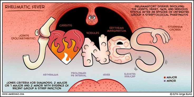
Acute rheumatic fever (ARF) is a sequella of symptoms that typically occur 2-4 weeks after an untreated bout of group A Streptoccocal (GAS) pharyngitis.  Symptoms include arthritis, carditis, erythema marginatum, CNS symptoms, and subcutaneous nodules.


Jones criteria is constellation of symptoms of ARF and are subdivided into major and minor manifestations.


Major

Carditis

Arthritis

CNS involvement

Subcutaneous nodules

Erythema marginatum

Minor

Arthralgia

Fever

Elevated acute phase reactants

Prolonged PR interval


The diagnosis of ARF is made using the Jones criteria and is positive if the patient has evidence of a preceding GAS infection and:

  • Two major manifestations

or

  • One major and two minor manifestations

How To Write A Personal Statement


This is a lecture I gave to a group of biomedical science students on how to write a personal statement for graduate professional programs.  It is directed towards medical professions (PA, MD, DO, DMD, etc.), but the basic tenets are the same.  By following a few basic rules and sprinkling in your unique experiences, your narrative will win over the selection committee.


To watch the video, click here


Infectious Disease Question


You are participating in a medical mission in South America and are seeing a 11yo boy who is brought in by his mother.  He has been complaining of joint pain and fever for the past 2 weeks.  She tells you that it seems to “move” from joint to joint over this time and nothing seems to help.  She does report that he has seemed to be sick for 4-6 weeks with various “cold” symptoms, but they didn’t seem too severe.  On examination, he has temperature of 101.2oF and the below rash.

erythema-marginatum-pictures-2


What should be your concern and what criteria can help make the diagnosis?

Update to Pneumonia Podcast


It figures.


As with any educational or academic en devour, as soon as you finish a paper/poster/presentation, a new publication comes out that would have been awesome to include.  I finished and published the Pneumonia podcast on July 12th and on July 14th, the IDSA/ATS released their updated guidelines on managing healthcare-associated (HAP) and ventilator-associated (VAP) pneumonia.  Luckily, they didn’t change anything earth-shattering, just tightened them up a bit.  Antibiotic regimens are below and they recommend 7-days total of therapy.

Screen Shot 2016-07-15 at 7.21.23 AM

2016 IDSA/ATS Guidelines

Screen Shot 2016-07-15 at 7.20.55 AM

2016 IDSA/ATS Guidelines


References

#16 – Pneumonia


***LISTEN TO THE PODCAST HERE***


Classifications of Pneumonia

  • Community-Acquired (CAP)
  • Healthcare-Associated (HAP)
    • Any IV therapy, wound care, or chemotherapy within 20 days
    • Resident of nursing home or other long term care facility
    • Hospitalization for ≥ 2 days within 90 days
    • Visit to outpatient clinic or hemodialysis within 30 days
  • Ventilator-Associated (VAP)
    • Currently or previously intubated during current hospitalization

Epidemiology

  • 6 cases per 1000 persons per year (~ 5 million cases per year)
  • Top 10 in mortality in US (~60,000 deaths/year)
  • 12% 30-day mortality in patients requiring admission
  • 28% all-cause mortality within one year

Pathogenesis

4 phases of development

  • Edema
    • Proteinaceous exudate in the alveoli
    • Bacteria accumulation
  • Red hepatization
    • Erythrocyte extravasation
  • Grey hepatization
    • Neutrophil extravasation with bacterial clearance
  • Resolution
    • Macrophage proliferation with inflammatory response
Picture1
Murthy SV. Pathology of Pneumonia. SlideShare.

Risk Factors

  • Age
  • Winter months
  • Increased risk of aspiration (AMS, CVA, intoxication, seizures)
  • Smoking
  • Underlying pulmonary disease (Asthma, COPD, cancer)
  • Immunosupression
  • Viral URI
  • Decreased host defenses (impaired ciliary clearance)
  • Acid-reducing medications
  • Malnutrition
  • Inhalation exposures
2
Up To Date. 2016.

Pathogens

  • Viral (most common)
    • Rhinovirus (most common)
    • Influenza
    • Adenovirus
    • Respiratory Syncytial Virus (RSV)
  • Bacterial
    • S. pneumoniae (most common CAP)
    • H. influenza
    • M. pneumoniae (most common atypical)
    • K. pneumoniae (tends to be more severe)
    • Legionella
    • ESKAPE bugs (>80% of VAP)
      • Enterococcus
      • Staphylococcus
      • Klebsiella
      • Acinetobacter
      • Pseudomonas
      • Enterobacter
  • Fungal (immunocompromised)
    • Histoplasmosis
    • Cryptococcus
    • Coccidioides
    • Blastomycosis
    • Aspergillus

Signs and Symptoms

  • Productive cough
  • Fever
  • Chills and/or rigors
  • Dyspnea
  • Pleuritic chest pain
  • Nausea/vomiting
  • Altered mental status

Physical Exam Findings

  • Vital signs
    • Febrile (elderly may not mount a response)
    • Tachycardic
    • Tachypnic
  • Pulmonary
    • Rales and/or rhonchi
    • Signs of consolidation
      • Decreased breath sounds
      • Dullness to percussion
      • Increased tactile fremitus
      • Egophany
      • Whispered pectoriliquoy

Radiographic Evaluation

  • Bacterial
    • Unilateral, lobar, air bronchograms
3
  • Viral
    • Diffuse or perihilar, bilateral
4

Laboratory Evaluation

  • CBC with differential
  • Blood cultures
  • Sputum culture and gram stain
    • Good sample = PMNs with < 10 squamous cells per LPF
  • Urine antigen (pneumococcal and Legionella)
  • Multiplex PCR
5
Up To Date. 2016.

Should They Stay or Should They Go Now????

  • Pneumonia Severity Index (PSI)
    • Step 1
      • If none of the following, then Class I and outpatient treatment
        • Age > 50 years
        • Neoplastic disease
        • Heart failure
        • Cerebrovascular disease
        • Renal disease
        • Liver disease
        • Altered mental status
        • HR ≥ 125/min
        • RR ≥ 30/min
        • SBP ≤ 90 mmHg
        • Temperature ≤ 35oC or ≥ 40oC
    • Step II
Screen Shot 2016-07-12 at 7.45.22 AM
Step II of PSI/PORT Score for Risk Stratification
  • CURB-65 Score
    • 5 variables
      • Confusion
      • Urea (BUN ≥ 20 mg/dL)
      • Respiratory rate ≥ 30/min
      • Blood pressure (SBP < 90 mmHg or DBP < 60 mmHg)
      • Age ≥ 65 years
    • Interpretation
      • Score 0-1 = Outpatient management
      • Score 2-3 = Inpatient management
      • Score 4-5 = ICU management
  • SMART-COP Score
    • Used to predict need for respiratory or vasopressor support
6

Treatment

  • Care should be taken to think about patients with risk factors for drug-resistant S. pneumoniae:
    • Age > 65 years
    • Beta-lactam, macrolide, or fluouroquinolone in the past 3-6 months
    • Alcoholism
    • Medical comorbidities
    • Immunosuppression
    • Exposure to child in daycare
  • Community-Acquired
7
Treatment for Community-Acquired Pneumonia. IDSA/ATS 2007 Guidelines.
  • Healthcare-Associated
8
Treatment of Healthcare-Associated Pneumonia. IDSA/ATS 2007 Guidelines.

Cottage Physician

CottageMD
Cottage Physician. 1893.

References

  1. Mandell LA, Wunderink RG. Pneumonia. In: Kasper D, Fauci A, Hauser S, Longo D, Jameson J, Loscalzo J. eds. Harrison’s Principles of Internal Medicine, 19e. New York, NY: McGraw-Hill; 2015. http://accessmedicine.mhmedical.com/content.aspx?bookid=1130&Sectionid=79733578. Accessed July 11, 2016.
  2. Guidelines for the management of adults with hospital-acquired, ventilator-associated, and healthcare-associated pneumonia. American journal of respiratory and critical care medicine. 171(4):388-416. 2005. [pubmed]
  3. File TM, Marrie TJ. Burden of community-acquired pneumonia in North American adults. Postgraduate medicine. 122(2):130-41. 2010. [pubmed]
  4. Murthy SV. Pathology of Pneumonia.    http://www.slideshare.net/vmshashi/pathology-of-pneumonia.  Accessed on July 11,  2016.
  5. Almirall J, Bolíbar I, Balanzó X, González CA. Risk factors for community-acquired pneumonia in adults: a population-based case-control study. The European respiratory journal. 13(2):349-55. 1999. [pubmed]
  6. Mandell LA, Wunderink RG, Anzueto A. Infectious Diseases Society of America/American Thoracic Society consensus guidelines on the management of community-acquired pneumonia in adults. Clinical infectious diseases : an official publication of the Infectious Diseases Society of America. 44 Suppl 2:S27-72. 2007. [pubmed]
  7. Musher DM, Thorner AR. Community-acquired pneumonia. The New England journal of medicine. 371(17):1619-28. 2014. [pubmed]
  8. Jain S, Self WH, Wunderink RG. Community-Acquired Pneumonia Requiring Hospitalization among U.S. Adults. The New England journal of medicine. 373(5):415-27. 2015. [pubmed]
  9. Metlay JP, Kapoor WN, Fine MJ. Does this patient have community-acquired pneumonia? Diagnosing pneumonia by history and physical examination. JAMA. 278(17):1440-5. 1997. [pubmed]
  10. Fine MJ, Auble TE, Yealy DM. A prediction rule to identify low-risk patients with community-acquired pneumonia. The New England journal of medicine. 336(4):243-50. 1997. [pubmed]
  11. Lim WS, van der Eerden MM, Laing R. Defining community acquired pneumonia severity on presentation to hospital: an international derivation and validation study. Thorax. 58(5):377-82. 2003. [pubmed]
  12. Charles PG, Wolfe R, Whitby M. SMART-COP: a tool for predicting the need for intensive respiratory or vasopressor support in community-acquired pneumonia. Clinical infectious diseases : an official publication of the Infectious Diseases Society of America. 47(3):375-84. 2008. [pubmed]
  13. Pugh R, Grant C, Cooke RP, Dempsey G. Short-course versus prolonged-course antibiotic therapy for hospital-acquired pneumonia in critically ill adults. The Cochrane database of systematic reviews. 2015. [pubmed]

Answer to Pulmonary Question

The diagnostic criteria for acute respiratory distress syndrome (ARDS) are the Berlin Criteria.

  1. Timing – within 7 days of a known clinical insult or worsening respiratory symptoms
  2. Imaging – Bilateral opacities not fully explained by effusions, atelectasis, or masses/nodules
  3. Absence of cardiac failure or fluid overload
  4. Hypoxemia – PaO2/FiO2 ≤ 300 mmHg with PEEP/CPAP ≥ 5 cmH2O


Screen Shot 2016-07-01 at 4.20.03 PM


Reference

  1. ARDS Definition Task Force.  Acute Respiratory Distress Syndrome – The Berlin Definition.  JAMA. 2012;307(23):2526-2533.
  2. PulmCCM Blog.  Meet the New ARDS: Expert panel announces new definition, severity classes.
  3. Life In The Fastlane Blog.  ARDS Definition.

#15 – Appendicitis


***LISTEN TO THE PODCAST HERE***


Epidemiology

  • 1st described in the late 1400s and 1st published in 1544 by Jean Fernal
  • 1st appendectomy was performed in 1736 on an 11yo boy
  • 233/100,000 population
  • May occur throughout life, but is most common age group is 10-19yo and slightly more common in men
  • Badass of the Millennium goes to….Leonid Rogozov.  He was the only physician on a Soviet team in Antarctica in 1961 and performed an appendectomy…on himself…and survived (Article here)

Picture1

Leonid Rogozov performing an appendectomy on himself – 1961


Anatomy of the Appendix

Wikipedia

Wikipedia

  • Located where the taenia coli converge at the base of cecum
  • Average length 6-9cm, outer diameter 3-8mm, and luminal diameter 1-3mm
  • Arterial supply is from appendicular branch of the ileocolic artery
  • The attachment of the base of the appendix is constant, but the tip may be positioned:
    • Retrocecal (most common)
    • Subcecal
    • Preileal
    • Postileal
    • Pelvic

Wikipedia

Wikipedia


Pathogenesis

  • Follows traditional predictable series of events for inflammation of a hollow visceral organ:
    • Inflammation
      • Most commonly by an obstruction
        • Fecalith, calculi, lymphoid hyperplasia, infection, mass
        • Causes a closed-loop obstruction
    • Distention
      • Normal secretion and bacterial overgrowth
        • Causes visceral nerve pain
      • Luminal pressure > perfusion
        • Involves the serosa
          • Causes parietal pain
    • Perforation
      • As areas of ischemia progress and pressures increase, perforation may occur
        • May be contained (localized) or cause peritonitis
      • Although this progression is predictable, the time frame may be variable
        • Perforation may occur anywhere from 24-48 hours after symptom onset

History

  • Abdominal pain occurs first
    • Starts as diffuse, periumbilical
    • Appendix position may alter pain location
  • Nausea and vomiting then follows
  • Anorexia (Negative Cheeseburger Sign)
  • Last is migratory pain to RLQ (50-60%)

Physical Examination

  • Often non-specific as appendix position, time course of illness, and patient anatomy may obscure findings
  • Classic physical exam findings include:
    • McBurney’s Point Tenderness
      • Maximal tenderness 3cm from ASIS, or 1/3rd the distance from ASIS to umbilicus
  • 4
    • Rovsing’s Sign
      • Palpation or rebound pressure of LLQ causes RLQ pain
    • Psoas Sign (retrocecal)
      • Pain in RLQ when ipsilateral hip is extended or flexed against resistance
    • Obsturator Sign (pelvic)
      • Pain in RLQ with internal rotation of ipsilateral hip with flexed knee
    • Guarding and peritoneal signs may be seen with perforation

5


Laboratory Studies

  • CBC with differential and CRP are needed for the scoring systems, but are not very specific

Pretest Probability Scoring Systems

  • Alvarado Score
    • Developed in 1986 and modified in 1994
  • Appendicitis Inflammatory Response (AIR) Score
    • Developed in 2008
  • Pediatric Appendicitis Score
    • Developed in 2002

Screen Shot 2016-06-15 at 12.39.04 PM

  • Interpretation
    • Alvarado Score
      • 0-3 – low probability (discharge)
      • 4-6 – indeterminate (image or admit)
      • ≥ 7 – high probability (admit or surgery)
    • AIR Score
      • 0-4 – Low probability (discharge)
      • 5-8 – Indeterminate (image or admit)
      • ≥ 9 – high probability (admit or surgery)
    • PAS
      • < 3 – low probability (discharge)
      • 3-7 – indeterminate (image or admit)
      • ≥ 8 – high probability (admit and consult)

Radiographic Imaging

  • Imaging is generally reserved for indeterminate cases or special populations (children, women, elderly) as clinical examination is just as effective as imaging in ruling-out appendicitis
  • Computed Tomography
    • IV contrast
    • Findings suggesting appendicitis:
      • ≥ 6mm diameter
      • Appendiceal wall thickening ≥ 2mm
      • Periappendiceal fat stranding
      • Appendicolith
  • Ultrasound
    • Study of choice in children and pregnant women and becoming study of choice in adults
    • Findings suggestive of appendicitis:
      • Aperistaltic, noncompressible, dilated (> 6mm) appendix
      • Distinct appendiceal wall layers
      • Echogenic prominent pericecal fat
      • Periappendiceal fluid collection
      • Target appearance on axial section

Metanalyses Comparing CT to US in Appendicitis

Metanalyses Comparing CT to US in Appendicitis


Management

  • Nonoperative Management
    • Several studies have looked at antibiotic management of uncomplicated appendicitis and found:
      • Up to 53% of antibiotic-only management still required surgery within 48hr
      • Antibiotic-only patients had lower pain scores and quicker return to work
      • Up to 37% of antibiotic-only patients required surgery for recurrent appendicitis within 2 years
      • Most recent study (APPAC Trial, JAMA, 2015) did meet prespecified criterion for noninferiority
      • May be an options for special populations
        • Poor surgical candidates, patients who refuse surgery
    • As for now, there isn’t enough good data to support routine, nonoperative management for uncomplicated appendicitis
  • Operative Management
    • Uncomplicated
      • Urgent (12-24hr) vs Emergent (< 12hr)
        • No difference in outcomes
        • 7
    • Complicated (perforation with abscess)
      • Sick or with generalized peritonitis = emergent surgery
      • Limited peritonitis
        • Antibiotics, bowel rest, percutaneous drainage
        • Interval appendectomy after improvement in clinical course
    • Laparoscopic vs Open Appendectomy
      • Laparoscopic > open in regards to patient outcomes
    • Complications
      • Wound infections
      • Organ space infections
      • Stump appendicitis

Cottage Physician

IMG_0305

Diagnosis and Management of Peritonitis


References

  1. Williams GR. Presidential Address: a history of appendicitis. With anecdotes illustrating its importance. Ann Surg. 1983;197(5):495-506.
  2. Buckius MT, Mcgrath B, Monk J, Grim R, Bell T, Ahuja V. Changing epidemiology of acute appendicitis in the United States: study period 1993-2008. J Surg Res. 2012;175(2):185-90.
  3. Liang MK, Andersson RE, Jaffe BM, Berger DH. The Appendix. In: Brunicardi F, Andersen DK, Billiar TR, Dunn DL, Hunter JG, Matthews JB, Pollock RE. eds. Schwartz’s Principles of Surgery, 10e. New York, NY: McGraw-Hill; 2014. http://accesssurgery.mhmedical.com/content.aspx?bookid=980&Sectionid=59610872. Accessed June 14, 2016.
  4. Raahave D, Christensen E, Moeller H, Kirkeby LT, Loud FB, Knudsen LL. Origin of acute appendicitis: fecal retention in colonic reservoirs: a case control study. Surg Infect (Larchmt). 2007;8(1):55-62.
  5. Birnbaum BA, Wilson SR. Appendicitis at the millennium. Radiology. 2000;215(2):337-48.
  6. Temple CL, Huchcroft SA, Temple WJ. The natural history of appendicitis in adults. A prospective study. Ann Surg. 1995;221(3):278-81.
  7. Andersson RE. Meta-analysis of the clinical and laboratory diagnosis of appendicitis. Br J Surg. 2004;91(1):28-37.
  8. McBurney C. Experience with early operative interference in cases of disease of the vermiform appendix.  NY State Med J. 1889;50:676.
  9. Alvarado A. A practical score for the early diagnosis of acute appendicitis. Ann Emerg Med. 1986;15(5):557-64.
  10. Kalan M, Talbot D, Cunliffe WJ, Rich AJ. Evaluation of the modified Alvarado score in the diagnosis of acute appendicitis: a prospective study. Annals of the Royal College of Surgeons of England. 76(6):418-9. 1994.
  11. Andersson M, Andersson RE. The appendicitis inflammatory response score: a tool for the diagnosis of acute appendicitis that outperforms the Alvarado score. World journal of surgery. 32(8):1843-9. 2008.
  12. De castro SM, Ünlü C, Steller EP, Van wagensveld BA, Vrouenraets BC. Evaluation of the appendicitis inflammatory response score for patients with acute appendicitis. World J Surg. 2012;36(7):1540-5.
  13. Samuel M. Pediatric appendicitis score. Journal of pediatric surgery. 37(6):877-81. 2002.
  14. Park JS, Jeong JH, Lee JI, Lee JH, Park JK, Moon HJ. Accuracies of diagnostic methods for acute appendicitis. The American surgeon. 2013;79(1):101-6.
  15. Whitley S, Sookur P, McLean A, Power N. The appendix on CT. Clinical radiology. 2009;64(2):190-9.
  16. Appendicitis. http://radiopaedia.org/articles/appendicitis. Accessed on June 14th, 2016.
  17. Sallinen V, Akl EA, You JJ. Meta-analysis of antibiotics versus appendicectomy for non-perforated acute appendicitis. Br J Surg. 2016;103(6):656-667.
  18. Varadhan KK, Neal KR, Lobo DN. Safety and efficacy of antibiotics compared with appendicectomy for treatment of uncomplicated acute appendicitis: meta-analysis of randomised controlled trials. BMJ. 2012;344:e2156.
  19. Salminen P, Paajanen H, Rautio T. Antibiotic Therapy vs Appendectomy for Treatment of Uncomplicated Acute Appendicitis: The APPAC Randomized Clinical Trial. JAMA. 2015;313(23):2340-8.
  20. Society of American Gastrointestinal and Endoscopic Surgeons. Guidelines for Laparoscopic Appendectomy.  http://www.sages.org/publications/guidelines/guidelines-for-laparoscopic-appendectomy/.  Accessed on June 15th, 2016.
  21. Blackborne LH. The Appendix. In: Surgical Recall. 6th Philadelphia, PA. LWW.