Ep-PAINE-nym


Bence Jones Protein

Other Known Aliasesurine monoclonal globulin protein

Definitionimmunoglobulin paraproteins produced by neoplastic plasma cells that are found in the urine due to decreased kidney filtration from acute kidney injury
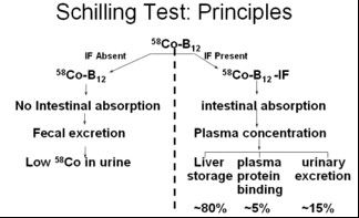
Clinical SignificanceBence Jones proteins are classically associated with multiple myeloma and Waldenström’s macroglobulinemia and these proteins were detected by heating a urine specimen to promote precipitation of the protein, but now is seen on electrophoresis of concentrated urine. Newer serum free light chain assays have been shown to be more sensitive and superior to the urine studies and are coming into favor.
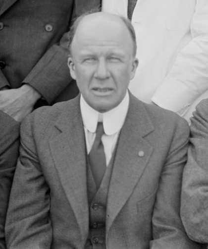
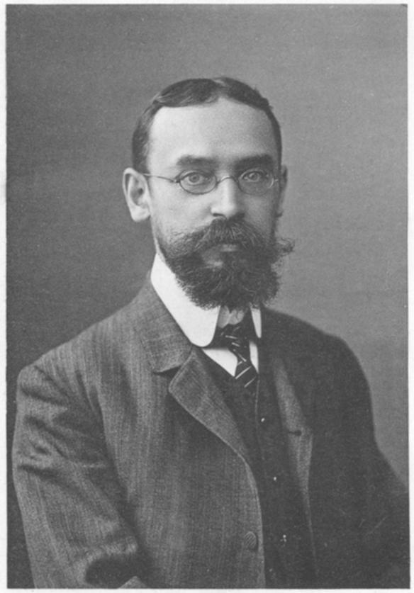
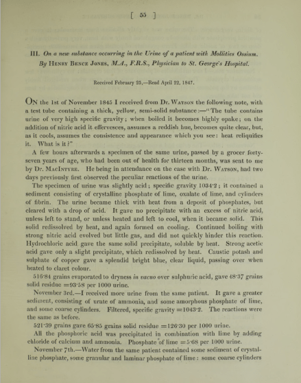
HistoryNamed after Henry Bence Jones (1813-1873), who was an English physician and chemist and received his medical doctorate from St. George’s Hospital in 1840. His love for chemistry was sparked during his medical training and he simultaneously undertook private instruction in chemistry studies from professor Thomas Graham. After medical school, he went to Giessen, Germany to train under Justus von Liebig’s (the leading chemist of his time) at his animalistic chemistry school. He described his eponymous finding in 1847 in an article entitled “On a new substance occurring in the urine of a patient with Mollities Ossium”. His work on applying chemistry principles to human disease was so far ahead of his time that his work was not nearly as successful as it should have been due to the lack of knowledge of biochemistry and physiology of the time.


References

  1. Firkin BG and Whitwirth JA.  Dictionary of Medical Eponyms. 2nd ed.  New York, NY; Parthenon Publishing Group. 1996.
  2. Bartolucci S, Forbis P.  Stedman’s Medical Eponyms.  2nd ed.  Baltimore, MD; LWW.  2005.
  3. Yee AJ, Pfiffner P. (2012).  Medical Eponyms (Version 1.4.2) [Mobile Application Software].  Retrieved http://itunes.apple.com.
  4. Whonamedit – dictionary of medical eponyms. http://www.whonamedit.com
  5. Up To Date. www.uptodate.com
  6. Katzmann JA, Abraham RS, Dispenzieri A, Lust JA, Kyle RA. Diagnostic performance of quantitative kappa and lambda free light chain assays in clinical practice. Clinical chemistry. 2005; 51(5):878-81. [pubmed]
  7. Jones HB. On a new substance occurring in the urine of a patient with mollities ossium. Philosophical Transactions of the Royal Society. 1848;138:55–62. doi:10.1098/rstl.1848.0003

Ep-PAINE-nym


Homan’s Sign

Other Known Aliasesdorsiflexion sign

Definitionpain in the posterior leg (classically behind the knee) with forced dorsiflexion of the foot

Clinical Significancethis examination finding was used in patients with a suspected DVT and before D-Dimers and clinical ultrasound were readily available. It is clinically useless as it has been studied extensively and found to have a sensitivity of 10-54% and specificity of 29-89%, thus not ruling in or out the condition consistently.

HistoryNamed after John Homans (1877-1954), who was an American surgeon and received his medical doctorate from Harvard Medical School. He worked with Harvey Cushing and Samuel Crowe early in career exploring the connection between the piuitary gland and the reproductive system. He first described his eponymous finding in 1944 in a NEJM article entitled “Diseases of the veins” and later published the first case report of a DVT occuring after prolonged sitting on a flight between Boston and Caracas in 1954. He was a founding member of the the Society for Vascular Surgery and the namesake of the John Homans Chair of Surgery position at Harvard Medical School and John Homans Fellowship in Vascular Surgery at the Brigham and Women’s Hospital.


References

  1. Firkin BG and Whitwirth JA.  Dictionary of Medical Eponyms. 2nd ed.  New York, NY; Parthenon Publishing Group. 1996.
  2. Bartolucci S, Forbis P.  Stedman’s Medical Eponyms.  2nd ed.  Baltimore, MD; LWW.  2005.
  3. Yee AJ, Pfiffner P. (2012).  Medical Eponyms (Version 1.4.2) [Mobile Application Software].  Retrieved http://itunes.apple.com.
  4. Whonamedit – dictionary of medical eponyms. http://www.whonamedit.com
  5. Up To Date. www.uptodate.com
  6. McGee, Steven (2012). Evidence-Based Physical Diagnosis. Philadelphia, USA: Saunders. pp. 472–473. ISBN978-1-4377-2207-9
  7. Homans J. Diseases of the veins. N Engl J Med 1944: 231; 51-60
  8. Homans J. Thrombosis of the deep leg veins due to prolonged sitting. The New England journal of medicine. 1954; 250(4):148-9. [pubmed]
  9. Barker WF. John Homans, MD, 1877-1954. Arch Surg. 1999;134(9):1019–1020. doi:10.1001/archsurg.134.9.1019

Ep-PAINE-nym


Schilling Test

Other Known Aliases – none

Definitionlaboratory test for pernicious anemia (specifically intrinisic factor deficiency) that led to vitamin B12 (cobalamin) deficiency. It involved ingesting a oral dose of radiolabeled vitamin B12 (to test oral absorption), an IM injection of vitamin B12 (to saturate liver stores), and a 24hr urine collection to see how much was absorbed and excreted. If intestinal absorption was intact (intrinsic factor present), then > 10% of the radiolabeled vitamin B12 would be in the urine.

Clinical SignificanceThis was the first and only test at the time to be able to diagnose pernicious anemia, but is now largely a test of historical interest only as better diagnostic studies have been developed.

HistoryNamed after Robert F. Schilling (1919-2014) an American physician and researcher who received his medical doctorate from the University of Wisconisn-Madison in 1943. Immediately after graduation, he joined the Pacific Front as a physician in the 3d Marine Division. After the war, he completed postgraduate training at Harvard before returning to Wisconsin to practice hematology. He studied extensively on the metabolism of vitamin B12 and the urinary excretion of radiolabeled vitamin B12 in pernicious anemia and in 1953, published a paper entitled “The effect of gastric juice on the urinary excretion of radioactivity after the oral administration of radioactive vitamin B12”, which would go on to be called the “Schilling Test”.


References

  1. Firkin BG and Whitwirth JA.  Dictionary of Medical Eponyms. 2nd ed.  New York, NY; Parthenon Publishing Group. 1996.
  2. Bartolucci S, Forbis P.  Stedman’s Medical Eponyms.  2nd ed.  Baltimore, MD; LWW.  2005.
  3. Yee AJ, Pfiffner P. (2012).  Medical Eponyms (Version 1.4.2) [Mobile Application Software].  Retrieved http://itunes.apple.com.
  4. Whonamedit – dictionary of medical eponyms. http://www.whonamedit.com
  5. Up To Date. www.uptodate.com
  6. Schilling RF. Intrinsic factor studies. 2. The effect of gastric juice on the urinary excretion of radioactivity after the oral administration of radioactive vitamin B12. J Clin Lab Med. 1953;42;860-866
  7. Schilling Test. Stat Pearls. 2018. https://www.ncbi.nlm.nih.gov/books/NBK507784/

Ep-PAINE-nym


Kruckenburg’s Tumor

Other Known Aliases – none

Definitionsecondary ovarian malignancy

Clinical SignificanceMost commonly arising from a gastric adenocarcinoma, but can occur from any metastatic cancer. 80% are bilateral and commonly manifest as pelvic pain, bloating, ascites, or dysparunea. Occasionaly, these tumor can be hormone producing and cause abnormal menstrual bleeding, hirsuitism, or virilization.

HistoryNamed after Friedrich Ernst Krukenberg (1871-1946), who was a German physician and received his medical doctorate from the University of Marburg.  He was actually studying to become a ophthalmologist, when he happend to be spending time in the pathology lab under Felix Marchand.  It was in this department that Krukenberg described a fibrosarcoma of the ovary (using sections from tumors Marchand had found in 1879) and published his findings in an article entitled “Über das Fibrosarcoma ovarii mucocellulare (carcinomatodes)” in 1896 at the age of 25 as part of his doctoral thesis. He spent the rest of career in his hometown of Halle, Germany practicing as a ophthalmologist.


References

  1. Firkin BG and Whitwirth JA.  Dictionary of Medical Eponyms. 2nd ed.  New York, NY; Parthenon Publishing Group. 1996.
  2. Bartolucci S, Forbis P.  Stedman’s Medical Eponyms.  2nd ed.  Baltimore, MD; LWW.  2005.
  3. Yee AJ, Pfiffner P. (2012).  Medical Eponyms (Version 1.4.2) [Mobile Application Software].  Retrieved http://itunes.apple.com.
  4. Whonamedit – dictionary of medical eponyms. http://www.whonamedit.com
  5. Up To Date. www.uptodate.com
  6. F. E. Krukenberg. Über das Fibrosarcoma ovarii mucocellulare (carcinomatodes).  Archiv für Gynäkologie. 1896;50:287-321.

Ep-PAINE-nym


Pfannensteil Incision

Other Known AliasesKerr incision

Definition8-10cm transverse (slightly arched) incision made 2-3cm cephalad to the pubic symphasis. The rectus sheath is then retracted cephalad and the rectus abdominis muscle bellies are divided longitudinally to enter the peritoneum

Clinical SignificanceThis is the primary incision for cesarean sections because it follows the Langer Lines and can achieve excellent cosmetic results. There are also decreased rates of postoperative pain, fascial dehiscence, and incisional hernias noted.

HistoryNamed after Hans Hermann Johannes Pfannensteil (1862-1909), who was a German gynecologist and received his medical doctorate from the University of Berlin in 1885. He was an extraordinary surgeon and teacher and published extensively on many gynecological conditions. In 1900, he published an article describing the the advantages of his eponymous transverse fascial incision for gynecologic laparotomies. Dr. Pfannensteil unfortunately died from septicemia at the age of 47 after injuring his finger draining a tubo-ovarian abscess.


References

  1. Firkin BG and Whitwirth JA.  Dictionary of Medical Eponyms. 2nd ed.  New York, NY; Parthenon Publishing Group. 1996.
  2. Bartolucci S, Forbis P.  Stedman’s Medical Eponyms.  2nd ed.  Baltimore, MD; LWW.  2005.
  3. Yee AJ, Pfiffner P. (2012).  Medical Eponyms (Version 1.4.2) [Mobile Application Software].  Retrieved http://itunes.apple.com.
  4. Whonamedit – dictionary of medical eponyms. http://www.whonamedit.com
  5. Up To Date. www.uptodate.com
  6. Pfannenstiel HJ. (On the advantages of the symphyseal transverse fascial incision for gynecological caliotomies as well as the contribution to the surgical indications). Samml Klin Vortr. 1900;268:1735-56

Ep-PAINE-nym


Leopold’s Maneuvers

Other known aliases – Leopold-Handgrïff

DefinitionSeries of four distinct actions to systematically determine the lie and position of the fetus in utero:

  • First Maneuver – Fundal Grip
    • used to locate fetal position (breech vs vertex)
  • Second Maneuver – Umbilical Grip
    • used to locate the back of the fetus
  • Third Maneuver – Second Pelvic Grip
    • used to determine pelvic inlet position
  • Fourth Maneuver – First Pelvic Grip
    • used to locate the fetal brow

Clinical SignificanceThese are now an antiquated way to determine fetal positioning to predict difficult deliveries or need for cesarean section. These have largely been replaced by obstetrical ultrasound.

HistoryNamed after Christian Gerhard Leopold (1846-1911), who was a German gynecologist and received his medical doctorate from the University of Leipzig in 1870. He spent the early part of his career teaching midwifery at the Frauenklinik in Leipiz before taking a professorship at the University of Leipzig in 1883. Later that same year, he took over as the Director of the Dresden Royal Gynaecological Infirmary and by the end of his tenure developed it into a leading hospital in Germany. He published his eponymous maneuvers in several articles (first in 1894) in the journal Archiv für Gynäkologie, for which he was a co-editor.


References

  • Firkin BG and Whitwirth JA.  Dictionary of Medical Eponyms. 2nd ed.  New York, NY; Parthenon Publishing Group. 1996.
  • Bartolucci S, Forbis P.  Stedman’s Medical Eponyms.  2nd ed.  Baltimore, MD; LWW.  2005.
  • Yee AJ, Pfiffner P. (2012).  Medical Eponyms (Version 1.4.2) [Mobile Application Software].  Retrieved http://itunes.apple.com.
  • Whonamedit – dictionary of medical eponyms. http://www.whonamedit.com
  • Up To Date. www.uptodate.com
  • Leopold CG – Die Leitung der regelmäßigen Geburt nur durch äußere Untersuchung. Arch Gynäkol. 1894; 45: 337–368
  • Kästner I, Kachlík D. German gynecologist and obstetrician Christian Gerhard Leopold (1846-1911). Ceska gynekologie. 2010; 75(3):218-21. [pubmed]

Ep-PAINE-nym


Roth’s Spots

Other know aliasesLitten’s spots

Definitionexudative, edematous hemorrhagic lesions of the retina with pale, white centers that can be composed of coagulated fibrin, platelets, infectious organisms, or neoplastic cells

Clinical Significanceone of the classic physical examination findings in bacterial endocarditis seen on fundoscopy. Further research and analysis has shown these can be present in leukemia, diabetes, and hypertensive retinopathy

Historynamed after Mortiz Roth (1839-1914), who was a Swiss pathologist and recieved his medical doctorate from University of Basel in 1864. He practiced all around Switzerland before returning to Basel as professor extraordinary of pathology in 1872, when he published his now eponymous findings in an article entitled “Uber Netzhauteffecstionen bei wundfiebren [Retinal Manifestations of wound fever]”. Dr. Roth, though, never described the classic appearance of the retinal red spot with a white center. Dr. Moritz Litten described this finding 6 years later and would coin the term we still use today.


References

  • Firkin BG and Whitwirth JA.  Dictionary of Medical Eponyms. 2nd ed.  New York, NY; Parthenon Publishing Group. 1996.
  • Bartolucci S, Forbis P.  Stedman’s Medical Eponyms.  2nd ed.  Baltimore, MD; LWW.  2005.
  • Yee AJ, Pfiffner P. (2012).  Medical Eponyms (Version 1.4.2) [Mobile Application Software].  Retrieved http://itunes.apple.com.
  • Whonamedit – dictionary of medical eponyms. http://www.whonamedit.com
  • Up To Date. www.uptodate.com
  • Roth Spots – StatPearls. [article]
  • Roth M. Uber Netzhauteffecstionen bei wundfiebren [Retinal manifestations of wound fever]. Deutsch A Chir. 1872;1:471–84.
  • Litten M. Ueber akute maligne endocarditis
  • und die dabei vorkommenden retinal veranderungen.
  • Charite-Ann 1878;3:135.

Ep-PAINE-nym


Korotkoff Sounds

Other known aliasesnone

Definitionsounds heard during auscultation of the brachial artery during blood pressure measurements

Clinical Significancethese sounds are generated as the sphygmomanometer cuff is slowly being deflated to the point that the maximal impulse of the pressure wave is more than cuff, but the cuff still occluded the artery at the nadir of the impulse. This pressure difference produces turbulence in the blood flow and the characteristic sound on auscultation. There are five phases to the Korotkov sounds with the initiation of Phase 1 as the systolic pressure and the end of Phase 4 as the diastolic pressure.

Historynamed after Nicholai Korotkov (1874-1920), who was a Russian surgeon and earned his medical degree from Moscow University in 1895. He had a prestigious career as a military physician and surgeon earning him an appointment as professor of surgery at the Military Medical Academy at St. Petersberg in 1903. He was preparing his doctoral thesis on vascular surgery when he described his now famous technique for measuring blood pressure in only a 281 word excerpt from a presentation to the Imperial Military Medical Academy entitled “Izvestie Imp. Voiennomedicinskoi Akademii” in 1905.

Nicolai Korotkov
Korotkov’s personal sphygmomanomter

References

  • Firkin BG and Whitwirth JA.  Dictionary of Medical Eponyms. 2nd ed.  New York, NY; Parthenon Publishing Group. 1996.
  • Bartolucci S, Forbis P.  Stedman’s Medical Eponyms.  2nd ed.  Baltimore, MD; LWW.  2005.
  • Yee AJ, Pfiffner P. (2012).  Medical Eponyms (Version 1.4.2) [Mobile Application Software].  Retrieved http://itunes.apple.com.
  • Whonamedit – dictionary of medical eponyms. http://www.whonamedit.com
  • Up To Date. www.uptodate.com
  • Shevchenko YL, Tsitlik JE. 90th Anniversary of the development by Nikolai S. Korotkoff of the auscultatory method of measuring blood pressure. Circulation. 1996; 94(2):116-8. [pubmed]

Ep-PAINE-nym


Bundle of Kent

Other known aliases atrioventricular bypass tract

DefinitionAs discussed in the WPW eponym, the Bundle of Kent is an accessory conduction pathway between the atrium and ventricle on either the right or left side of the heart.

Clinical Significancethis pathway occurs in up to 0.3% of patients and the cause of Wolff-Parkinson-White syndrome. It bypasses the traditional conduction system and allows for pre-excitation tachydysrthymias.

HistoryNamed after Albert Frank Stanley Kent (1863-1958), an English physiologist who received his degree in 1886 from the Magdalen College of Oxford. He first described lateral atrioventricular connections in a monkey heart in 1893 and erroneously believed these were part of the normal specialized conduction system. These findings generated a lot of controversy at the time and were actually rejected by several notable anatomists and physiologists. In fact, in 1955, Lev and Learner dissected 33 neonatal hearts and found no evidence of “normal” lateral conduction systems.


References

  • Firkin BG and Whitwirth JA.  Dictionary of Medical Eponyms. 2nd ed.  New York, NY; Parthenon Publishing Group. 1996.
  • Bartolucci S, Forbis P.  Stedman’s Medical Eponyms.  2nd ed.  Baltimore, MD; LWW.  2005.
  • Yee AJ, Pfiffner P. (2012).  Medical Eponyms (Version 1.4.2) [Mobile Application Software].  Retrieved http://itunes.apple.com.
  • Whonamedit – dictionary of medical eponyms. http://www.whonamedit.com
  • Up To Date. www.uptodate.com
  • Kent AF. Researches on the Structure and Function of the Mammalian Heart. The Journal of physiology. 1893; 14(4-5):i2-254. [pubmed]
  • LEV M, LERNER R. The theory of Kent; a histologic study of the normal atrioventricular communications of the human heart. Circulation. 1955; 12(2):176-84. [pubmed]

Ep-PAINE-nym


Wolff-Parkinson-White Syndrome

Other known aliasesventricular pre-excitation with arrhythmia, auriculoventricular accessory pathway syndrome

Definitionparoxysmal supraventricular tachycardia caused by conduction through an abnormal accessory bypass tract between the atria and ventricles known as the Bundle of Kent. There are two types depending on the side of the heart it effects; Type A is between the right atrium and ventricle and Type B is between the left atrium and ventricle.

Clinical SignificancePatients with WPW can numerous cardiac dysfunction symptoms including tachydysrhythmias, palpitations, dyspnea, presyncope, syncope, and sudden cardiac arrest. It is characterized by the triad of abnormalities on EKG of widened QRS, shortened PR interval, and slurring of the initial part of the QRS (called a delta wave).

HistoryNamed after Louis Wolff (1898-1972), Sir John Parkinson (1885-1976), and Paul Dudley White (1886-1973). Dr. Wolff was an American cardiologist who received his medical doctorate from Harvard Medical School in 1922. Dr. Parkinson was an English cardiologist who received his medical doctorate from University of Freiburg in 1910 and was also knighted by King George in 1948. Dr. White was an American cardiologist who received his medical doctorate from Harvard Medical School in 1911 and one of the founding presidents for the American Heart Association. He was a prominent advocate for preventive medicine receiving many national and international awards for his efforts to advance the importance of diet, exercise, and weight control in the prevention of cardiovascular disease. They collaborated to publish a series of 11 cases entitled “Bundle‐Branch Block with Short P‐R Interval in Healthy Young People Prone to Paroxysmal Tachycardia” in the American Heart Journal in 1930. It should be noted that Dr. Frank Norman Wilson and Dr. Alfred Wedd both described and published these findings in 1915 and 1921.


References

  • Firkin BG and Whitwirth JA.  Dictionary of Medical Eponyms. 2nd ed.  New York, NY; Parthenon Publishing Group. 1996.
  • Bartolucci S, Forbis P.  Stedman’s Medical Eponyms.  2nd ed.  Baltimore, MD; LWW.  2005.
  • Yee AJ, Pfiffner P. (2012).  Medical Eponyms (Version 1.4.2) [Mobile Application Software].  Retrieved http://itunes.apple.com.
  • Whonamedit – dictionary of medical eponyms. http://www.whonamedit.com
  • Up To Date. www.uptodate.com
  • Wolff L, Parkinson J, White PD. Bundle‐Branch Block with Short P‐R Interval in Healthy Young People Prone to Paroxysmal Tachycardia. American Heart Journal. 1930;5(6):985-704 [article]