#41 – Bell’s Palsy


***LISTEN TO THE PODCAST HERE***


Anatomy and Physiology of Cranial Nerve VII

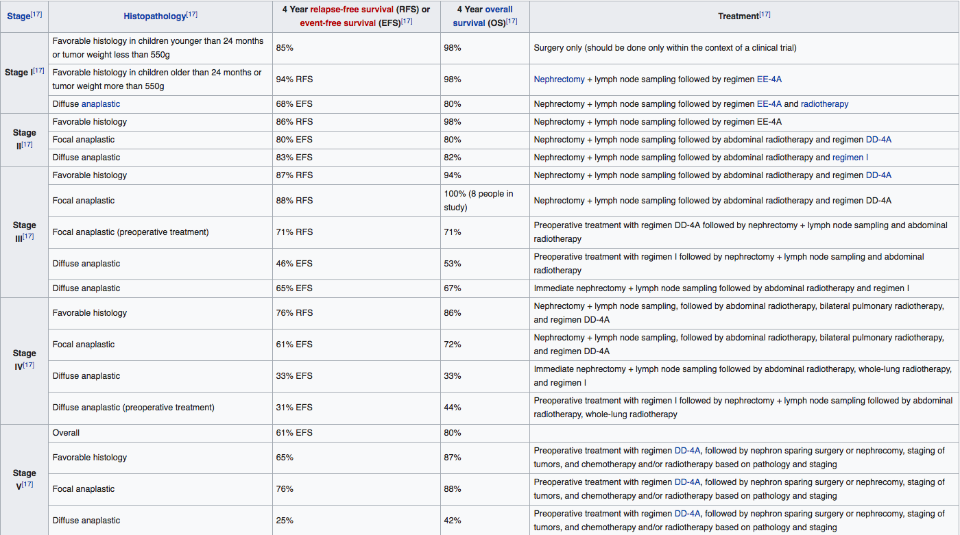
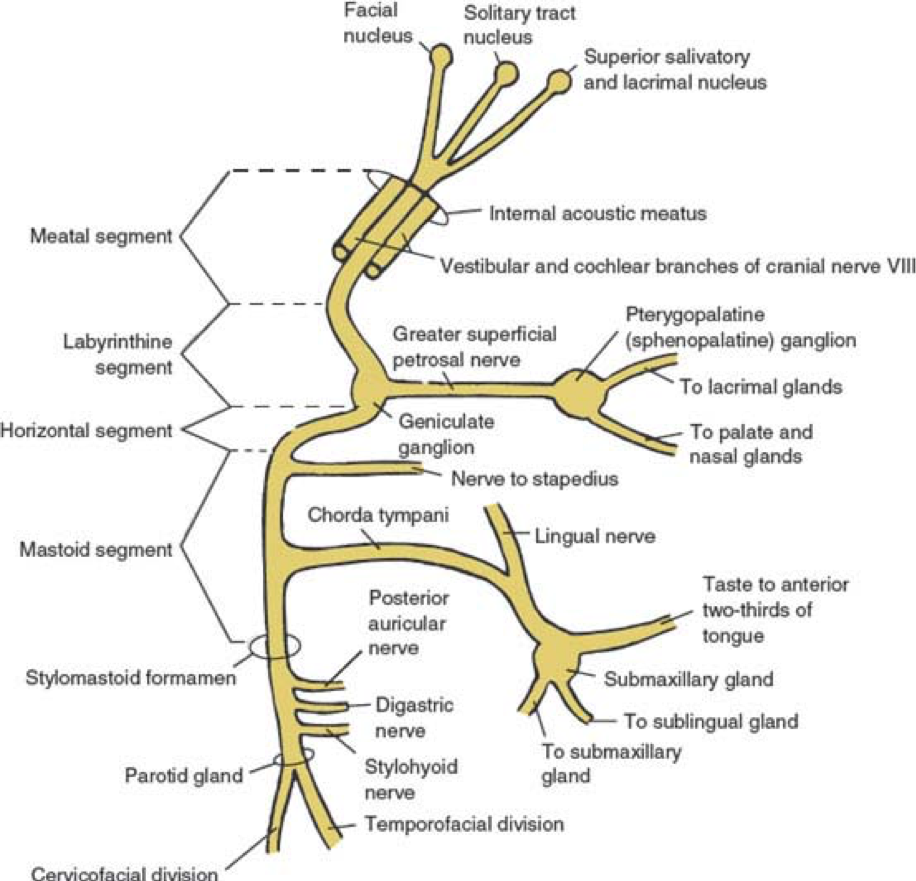
Cranial Nerve VII (the facial nerve) is a mixed nerve (having both sensory and motor function) with:

  • Somatic afferents from the external auditory canal
  • Afferent fibers from taste receptors from the anterior 2/3rds of the tongue
  • Parasympathetic fibers innervating lacrimal, submandibular, and sublingual salivary glands
  • Motor fibers innervating the facial muscles

There are six (6) segments of the nerve after it emerges from the pons of the brainstem:

  1. Intracranial (cisternal)
  2. Meatal (from brainstem to internal auditory canal)
  3. Labyrinthine (internal auditory canal to geniculate ganglion)
    1. Lacrimal gland, nasal/palate, pterygopalatine ganglion
  4. Tympanic (geniculate ganglion to pyramidal eminence)
    1. Stapedius nerve
  5. Mastoid (pyramidal eminence to stylomastoid foramen)
    1. Chorda tympani à tongue, salivary glands, lingual nerve
  6. Extratemporal (stylomastoid foramen to post parotid branches)
    1. 5 major facial branches
      1. Temporal
      2. Zygomatic
      3. Buccal
      4. Mandibular
      5. Cervical


Definition and Epidemiology

Bell’s Palsy is an acute peripheral nerve palsy of unknown etiology and makes up almost half of such cases.  It is estimated that the annual incidence is around 20 patient per 100,000 population.  There is no race, geographic, or gender correlation., though there is some old data on increased risk during 3rd trimester of pregnancy.


Pathogenesis

Had been hotly debated for many years as to the cause of this condition and is generally considered to be caused by Herpes Simplex virus due to associated serologic evidence in effected patients.  Newer data has been published, but is not entirely conclusive.  Most agree that is due to some viral pathogen with herpes being the most common, but also implicating CMV, EBV, adenovirus, and coxsackievirus. Specifically, the signs and symptoms are a result of viral mediated inflammatory demyelination of the nerve.


Signs and Symptoms

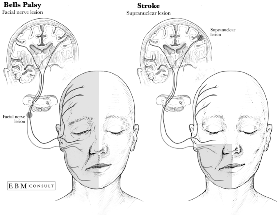
Most common presentation is acute (over several hours) unilateral facial paralysis with:

  • Motor
    • Inability to close the eye
    • Eyebrow sagging with inability to wrinkle the forehead
    • Obliteration of the nasolabial fold
    • Dropping of the affected corner of the mouth
  • Sensory
    • Hyperacusis
    • Loss of taste on the anterior 2/3rd of tongue

Symptoms are progressive and usually reach peak involvement within 3 weeks of onset.  Patients should begin to have return of function within 2-4 months.


Diagnostic Studies

Bell’s palsy is a clinical diagnosis and diagnostic studies are generally not indicated unless the presentation is atypical, still progressing at 3 weeks, or there is no return of function by 4 months.  History of facial twitching or spasms preceding the paralysis raises suspicion of compressive neuropathy from tumor or mass.

  • Electromyography (EMG)
    • The most simplest of the electrodiagnostic tests that can be used to show action potentials on active volition.  Some degree of potential infers that the nerve is still intact and therefor, can improve.
  • Nerve Conduction Studies (NCS)
    • Supramaximal stimulation near the parotid gland with measured evoked potentials over the orbicularis oculi, nasalis, and lower facial muscles  can measure the degree of axonal loss.  Studies have shown >75% is the critical cutoff for low likelihood of full recovery.  Ideally, this should be performed within 2 weeks of symptom onset for a more accurate prognosis.
  • Facial Nerve Stimulation
    • Should be considered with 2 weeks of symptoms onset if surgical decompression is considered due to potential for reversibility.
  • Needle EMG
    • Utilized after 3 weeks to assess the degree of axonal damage and evidence of subclinical reinnervation for recovery
  • Imaging
    • High-resolution, contrast-enhanced CT (for bony pathology) or gadolinium-enhanced MRI (for soft tissue structures) of the brain, temporal bone, and parotid gland.
  • Lyme serology should be entertained in patients from endemic regions.
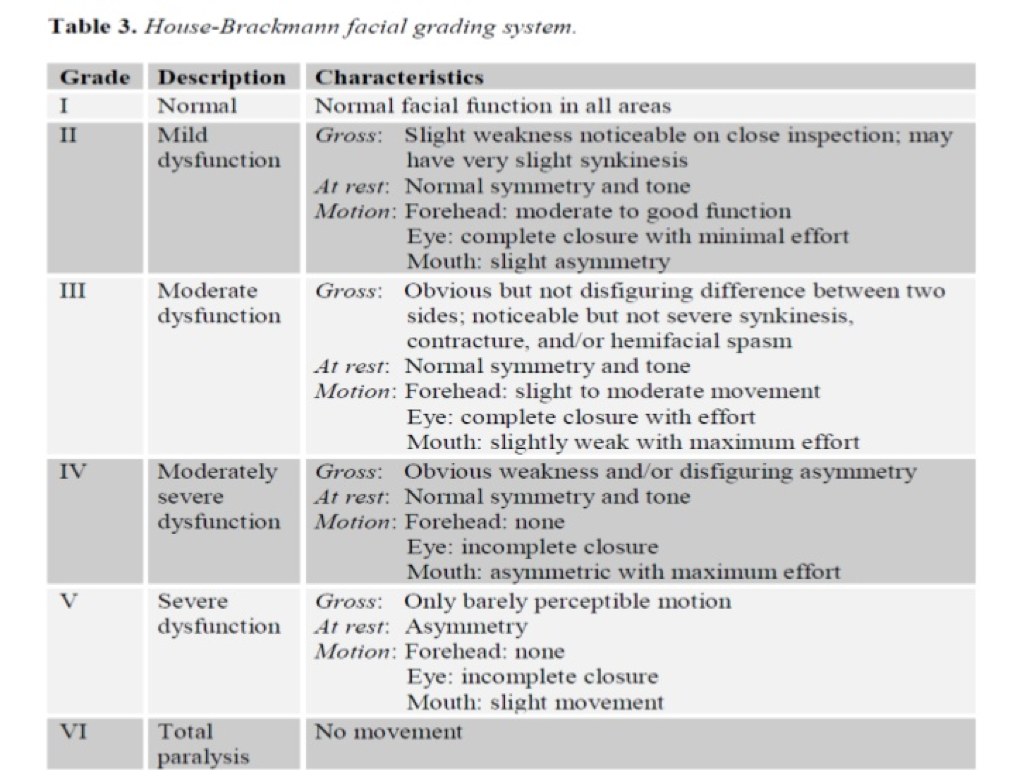
Severity Grading

The House-Brackman scale is the most common grading scale that uses objective criteria to score the severity, mark progression, and track return of function.


Treatment

Mainstay of therapy for Bell’s palsy is corticosteroids (for the inflammation) and antivirals (for the viral pathogens).  Current recommendations are:

  • Prednisone 60-80mg daily for 7 days
  • Valacyclovir 1000mg TID for 7 days
  • Within 72 hours of symptoms onset


Eye care should include artificial tears and an eye patch for sleeping.


Prognosis

Favorable prognosis if any recovery is seen within 21 days of symptom onset with data showing 71% with complete resolution, 13% with slight residual sequelae, and 16% with residual weakness, synkinesis, or contracture.  94% of patients with incomplete involvement have full resolution, while only 60% of complete involvement returned to normal function.


References

  1. Monkhouse WS. The anatomy of the facial nerve. Ear, nose, & throat journal. 1990; 69(10):677-83, 686-7. [pubmed]
  2. May M, Klein SR. Differential diagnosis of facial nerve palsy. Otolaryngologic clinics of North America. 1991; 24(3):613-45. [pubmed]
  3. Hilsinger RL, Adour KK, Doty HE. Idiopathic facial paralysis, pregnancy, and the menstrual cycle. The Annals of otology, rhinology, and laryngology. ; 84(4 Pt 1):433-42. [pubmed]
  4. Peitersen E. Bell’s palsy: the spontaneous course of 2,500 peripheral facial nerve palsies of different etiologies. Acta oto-laryngologica. Supplementum. 2002; [pubmed]
  5. Schirm J, Mulkens PS. Bell’s palsy and herpes simplex virus. APMIS : acta pathologica, microbiologica, et immunologica Scandinavica. 1997; 105(11):815-23. [pubmed]
  6. Kennedy PG. Herpes simplex virus type 1 and Bell’s palsy-a current assessment of the controversy. Journal of neurovirology. 2010; 16(1):1-5. [pubmed]
  7. Morgan M, Nathwani D. Facial palsy and infection: the unfolding story. Clinical infectious diseases : an official publication of the Infectious Diseases Society of America. 1992; 14(1):263-71. [pubmed]
  8. Liston SL, Kleid MS. Histopathology of Bell’s palsy. The Laryngoscope. 1989; 99(1):23-6. [pubmed]
  9. Valls-Solé J. Electrodiagnostic studies of the facial nerve in peripheral facial palsy and hemifacial spasm. Muscle & nerve. 2007; 36(1):14-20. [pubmed]
  10. May M, Klein SR, Taylor FH. Indications for surgery for Bell’s palsy. The American journal of otology. 1984; 5(6):503-12. [pubmed]
  11. Gilden DH. Clinical practice. Bell’s Palsy. The New England journal of medicine. 2004; 351(13):1323-31. [pubmed]
  12. House JW, Brackmann DE. Facial nerve grading system. Otolaryngology–head and neck surgery : official journal of American Academy of Otolaryngology-Head and Neck Surgery. 1985; 93(2):146-7. [pubmed]
  13. Gronseth GS, Paduga R, . Evidence-based guideline update: steroids and antivirals for Bell palsy: report of the Guideline Development Subcommittee of the American Academy of Neurology. Neurology. 2012; 79(22):2209-13. [pubmed]
  14. Baugh RF, Basura GJ, Ishii LE, et al. Clinical practice guideline: Bell’s palsy. Otolaryngology–head and neck surgery : official journal of American Academy of Otolaryngology-Head and Neck Surgery. 2013; 149(3 Suppl):S1-27. [pubmed]
  15. Schwartz SR, Jones SL, Getchius TS, Gronseth GS. Reconciling the clinical practice guidelines on Bell’s palsy from the AAO-HNSF and the AAN. Otolaryngology–head and neck surgery : official journal of American Academy of Otolaryngology-Head and Neck Surgery. 2014; 150(5):709-11. [pubmed]
  16. de Almeida JR, Guyatt GH, Sud S, et al. Management of Bell palsy: clinical practice guideline. CMAJ : Canadian Medical Association journal = journal de l’Association medicale canadienne. 2014; 186(12):917-22. [pubmed]
  17. Peitersen E. The natural history of Bell’s palsy. The American journal of otology. 1982; 4(2):107-11. [pubmed]

PAINE #PANCE Pearl – Neurology


Questions


  1. What is the popular scoring system for determining a patient’s need for anticoagulation to prevent stroke in atrial fibrillation?
  2. If you do start anticoagulation, what are some scoring systems to determine a patient’s risk for bleeding while on anticoagulation?


Image result for atrial fibrillation stroke

Image result for atrial fibrillation stroke

Ep-PAINE-nym


Klinefelter Syndrome


Other Known Aliases47XXY, Klinefelter-Reifenstein-Albright Syndrome


DefinitionGenetic condition resulting from two (or more) X chromosomes in a male patient.  It is the most common sex chromosome abnormality causing hypogonadism.

Human chromosomesXXY01.png

Clinical SignificanceThis condition affects 1 in 500-1000 newborn males in the United States.  Symptoms can range from subtle (sometimes not even noticed) to severe learning, developmental, and cognitive deficiencies.  The most prominent features are sterility, small testes, taller stature, less androgenic body hair, and gynecomastia.  Due to these developmental abnormalities, it is often not diagnosed until after puberty

Image result for klinefelter syndrome

History – Named after Harry Fitch Klinefelter, Jr. (1912-1990), an American rheumatologist and endocrinologist, who earned is medical doctorate from Johns Hopkins University.  He worked in the prestigious clinic of Dr. Fuller Albright at Massachusetts General Hospital where he studied a group of nine boys who all had similar features of gynecomastia, aspermatogenesis, and increased follicle-stimulating hormone.  He credits Dr. Albright with this discovery, as it was his clinic and he first noticed the pattern, but Albright wanted Klinefelter to do the research work on it as a new fellow attending.  The group (with Dr. Edward Reifenstein) published this cases series in 1942 and Dr. Albright insisted that Klinefelter take lead authorship.


Image result


References

  1. Firkin BG and Whitwirth JA.  Dictionary of Medical Eponyms. 2nd ed.  New York, NY; Parthenon Publishing Group. 1996.
  2. Bartolucci S, Forbis P.  Stedman’s Medical Eponyms.  2nd ed.  Baltimore, MD; LWW.  2005.
  3. Yee AJ, Pfiffner P. (2012).  Medical Eponyms (Version 1.4.2) [Mobile Application Software].  Retrieved http://itunes.apple.com.
  4. Whonamedit – dictionary of medical eponyms. http://www.whonamedit.com
  5. Up To Date. www.uptodate.com
  6. Klinefelter Syndrome.  https://ghr.nlm.nih.gov/condition/klinefelter-syndrome
  7. Klinefelter HF, Reifenstein EC, Albright F. Syndrome Characterized by Gynecomastia, Aspermatogenesis without A-Leydigism, and Increased Excretion of Follicle-Stimulating Hormone.  JCEM.  1942;2(11):615-627 [article]
  8. Loriau DL.  Chapter 89: Harry F. Klinefelter (1912-1990).  A Biographical History of Endocrinology. 2016.  [article]

PAINE #PANCE Pearl – Pediatrics


Question


A 3yo girl has been diagnosed with acute bacterial otitis media in your clinic and requires amoxicillin for treatment.  The parents say she doesn’t take medicine every well and would appreciate the lowest VOLUME per dose.  She weighs 32 lbs.  What are the different dosing strategies using the current formulation of liquid amoxicillin?


Answer


Amoxicillin comes in 125mg/5mL, 200mg/5mL, 250mg/5mL, and 400mg/5mL and the recommended daily dose for bacterial acute otitis media is 90mg/kg/day twice daily for 7 days. She weighs 32lbs, which is 14.5kg, so she would need 1305mg per day, or 650mg per dose. For the different concentrations it could be:

  • For 125mg/5mL – 26mL per dose
  • For 200mg/5mL – 16mL per dose
  • For 250mg/5mL – 13mL per dose


If you want to have the LOWEST volume, then using the 400mg/5mL concentration would be 8.1mL per dose and you would need to dispense 115mL for a 7 day prescription.


Ep-PAINE-nym


Wilms Tumor


Other Known Aliasesnephroblastoma


DefinitionThe most common childhood primary renal tumor and can occur due to a mutation in the WT-1 cancer suppressor gene on 11p13.  There are five clinical stages depending on anatomical findings and tumor cell pathology.


Image result for wilms tumor


Clinical SignificanceCurrent estimates are around 500 new cases in the US per year and can be associated with several other genetic conditions including, WAGR syndrome, Denys-Drash syndrome, and Beckwith-Wideman syndrome.   Most children, who are later diagnosed with a Wilm’s tumor, present with an asymptomatic abdominal mass easily palpable by the provider.   Ultrasound is the initial screening test of choice, though MRI can be help in staging.  A renal biopsy will confirm the diagnose and definitively stage the disease in order to select the best treatment modalities.


History – Named after Carl Max Wilhem Wilms (1867-1918), a German pathologist and surgeon,  who earned his medical doctorate from the University of Bonn in 1890.  He was a prolific surgeon and medical educator rising to the ranks of chair of surgery at the University of Heidelberg in 1910.  He published his findings of the renal tumor that bears his name in 1899 in the article entitled “Die Mischgeschwülste der Niere”.  There is some controversy on who identified this tumor first as Thomas Rance may have written on it in 1814, but it was not very specific and could be attributed to other renal malignancies.   Felix Birch-Hirschfeld and colleagues also identified and wrote on what they believed to the first description of this tumor, but Wilms’ manuscript seemed to be more broadly noted in the literature and eventually came to bear his name.  


Image result for Die Mischgeschwülste der Niere


References

  1. Firkin BG and Whitwirth JA.  Dictionary of Medical Eponyms. 2nd ed.  New York, NY; Parthenon Publishing Group. 1996.
  2. Bartolucci S, Forbis P.  Stedman’s Medical Eponyms.  2nd ed.  Baltimore, MD; LWW.  2005.
  3. Yee AJ, Pfiffner P. (2012).  Medical Eponyms (Version 1.4.2) [Mobile Application Software].  Retrieved http://itunes.apple.com.
  4. Whonamedit – dictionary of medical eponyms. http://www.whonamedit.com
  5. Wilm’s Tumor.  National Cancer Institute. https://www.cancer.gov/types/kidney/hp/wilms-treatment-pdq#link/_1
  6. Up To Date. Accessed April 21, 2018.  www.uptodate.com
  7. Coppes-Zantingal AR.  Max Wilms and “Die mischgeschwülste der Niere”.  CMAJ.  199;160(8):1196. [pubmed]
  8. Coppes AR.  Dr. Carl Max Wilhelm Wilms.  HemOnc Today.  2008.  https://www.healio.com/hematology-oncology/pediatric-oncology/news/print/hemonc-today/%7B05646a17-7a0c-4a39-a0db-62fd83e0d628%7D/dr-carl-max-wilhelm-wilms-1867-1918

PAINE #PANCE Pearl – Pediatrics


Question


A 3yo girl has been diagnosed with acute bacterial otitis media in your clinic and requires amoxicillin for treatment.  The parents say she doesn’t take medicine every well and would appreciate the lowest VOLUME per dose.  She weighs 32 lbs.  What are the different dosing strategies using the current formulation of liquid amoxicillin?


Ep-PAINE-nym


Crigler-Najjar Syndrome


Other Known Aliasesfamilial/hereditary nonhemolytic unconjugated hyperbilirubinemia


DefinitionRare, familial condition resulting in congenital hyperbilirubinemia secondary to a deficiency of glucuronyl transferase.  There two types, with type I being very rare and severe (absolute absence) and type II being more common and less severe (relative deficiency)

Image result for bilirubin metabolism


Clinical Significance – Congenital hyperbilirubinemia can have catastrophic effects on the infant including jaundice, lethargy, failure to thrive, hypotonia, kernicterus, and acute bilirubin encephalopathy once it saturates and binds to the brain tissue.


History – Named after:

1) John Fielding Crigler (1919-), who is an American pediatrician who earned his medical doctorate at Duke University in 1943 and practiced at the Children’s Hospital of Boston.

2) Victor Assad Najjar (1914-), who is a Lebonese-born, American pediatrician who earned his medical doctorate at the American University in Beirut and practiced at Johns Hopkins, Vanderbilt, and Tufts University.


They published their findings of a new disease causing congenital familial nonhemolytic jaundice with kernicterus in 1952.


References

  1. Firkin BG and Whitwirth JA.  Dictionary of Medical Eponyms. 2nd ed.  New York, NY; Parthenon Publishing Group. 1996.
  2. Bartolucci S, Forbis P.  Stedman’s Medical Eponyms.  2nd ed.  Baltimore, MD; LWW.  2005.
  3. Yee AJ, Pfiffner P. (2012).  Medical Eponyms (Version 1.4.2) [Mobile Application Software].  Retrieved http://itunes.apple.com.
  4. Whonamedit – dictionary of medical eponyms. http://www.whonamedit.com
  5. Crigler-Najjar Syndrome.  Genetics Home Reference. https://ghr.nlm.nih.gov/condition/crigler-najjar-syndrome#synonyms
  6. CRIGLER JF, NAJJAR VA. Congenital familial nonhemolytic jaundice with kernicterus; a new clinical entity. A.M.A. American journal of diseases of children. 1952; 83(2):259-60. [pubmed]

PAINE #PANCE Pearl – Pediatrics


Question


Image result for omphaloceleImage result for gastroschisis


  1. What are these two conditions called?
  2. Which is associated with other genetic conditions?
  3. Which has better associated outcomes?


Answer


  1. The condition on the left is an omphalocele and on the right is a gastroschisis.
    1. Omphalocele is a midline abdominal wall defect where the contents are covered by a membrane of amnion and peritoneum.  It occurs at the base of umbilical cord.
    2. Gastroschisis (more common) is a full-thickness, paraumbilical abdominal wall defect with free-evisceration of abdominal contents
  2. The majority of infants with an omphalocele have associated congenital anomalies including Beckwith-Wiedemann syndrome, Trisomy 13 and 18, and numerous other subsystem involvement.  Gastroschisis generally has no other abnormalities, but can have associated intestinal atresia in up to 10%
  3. Gastroschisis has statistically better overall outcomes since it is not associated with any other genetic conditions.

Image result for omphalocele vs gastroschisis


References

  1. Carlo WA.  The Umbilicus. In: Nelson Textbook of Pediatrics. 20th ed.  2016. Chapter 105: 890-891.
  2. Abdominal Wall Defect.  Genetics Home Reference.  https://ghr.nlm.nih.gov/condition/abdominal-wall-defect

Ep-PAINE-nym


Kayser-Fleischer Rings


Other Known Aliasesnone


Definition1-3mm, grey/green/brown pigmented ring in the Descemet membrane of the cornea.  It first appears at the 12 o’clock position in early disease, then a second crescent forms at 6 o’clock, and then finally completely encircling the cornea.


Kayser-Fleischer ring.jpg


Clinical Significance – This is pathognomonic for Wilson’s disease, but does not cause any symptoms with the patient.  They are often identified on good ophthalmic examinations.


History – Named after Bernhard Kayser (1869-1954) and Bruno Fleischer (1874-1965), who were both German ophthalmologists and contemporaries of each other.  Dr. Kayser received his medical doctorate at the University of Berlin in 1893 and practiced as a specialized ophthalmologist in Stuttgart, Germany for the majority of his career.  Dr. Fleischer received his medical doctorate at the University of Tübingen in 1898 and practiced there earning a reputation as an extraordinary professor.  Each published their findings in Klinische Monatsblätter für Augenheilkunde within a year of each other (1902 and 1903), but erroneously posited that it was due to silver accumulation.  The first published report of copper being the causative agent was in 1934 by Dr. Werner Gerlach and Willhelm Rohrschneider.


References

  1. Firkin BG and Whitwirth JA.  Dictionary of Medical Eponyms. 2nd ed.  New York, NY; Parthenon Publishing Group. 1996.
  2. Bartolucci S, Forbis P.  Stedman’s Medical Eponyms.  2nd ed.  Baltimore, MD; LWW.  2005.
  3. Yee AJ, Pfiffner P. (2012).  Medical Eponyms (Version 1.4.2) [Mobile Application Software].  Retrieved http://itunes.apple.com.
  4. Whonamedit – dictionary of medical eponyms. http://www.whonamedit.com
  5. Schrag A, Schott JM. Images in clinical medicine. Kayser-Fleischer rings in Wilson’s disease. NEJM. 2012; 366(12):e18. [pubmed]
  6. Dusek P, Litwin T, Czlonkowska A. Wilson disease and other neurodegenerations with metal accumulations. Neurologic clinics. 2015; 33(1):175-204. [pubmed]
  7. Kayser B. “Über einen Fall von angeborener grünlicher Verfärbung des Cornea”. Klin Monatsbl Augenheilk. 1902;40(2):22–25.
  8. Fleischer B. “Zwei weitere Fälle von grünlicher Verfärbung der Kornea”. Klin Monatsbl Augenheilk. 1903;41(1):489–491
  9. Gerlach W, Rohrschneider W. “Besteht das Pigment des Kayser-Fleischerschen Hornhautringes aus Silber?”. Klin Wochenschr. 1934;13: 48–49

#40 – Well Child Visits


***LISTEN TO THE PODCAST HERE***


2015 American Academy of Pediatrics Schedule for Preventive Pediatric Health


2018 Centers for Disease Control Immunization Schedule


Denver Developmental Milestones


Initial Office Visit (within 2 weeks)


Development

  • Rooting and suckling reflex strong
  • Indiscriminate tight grasp of hands
  • Responds to light, noise, and movement
  • Lift head from supine
  • Follow to midline
  • Regained birthweight


Anticipatory Guidance

  • Feeding
    • Encourage breastfeeding and its benefits:
      • Decreased risk of infections
      • Decreased SIDS risk
      • Decreased obesity risk
      • Increases emotional connection
    • Frequency
      • On-demand, but generally every 2-4 hours
    • Amount
      • 1 oz/kg/day
    • Reflux
  • Sleeping
    • Back to sleep
  • Fever
  • Jaundice

Vaccinations

  • Hepatitis B #1 (if not given in hospital)


2 Month Visit


Development

  • Smile socially and start cooing
  • Lift head from prone
  • Turns head towards sounds and follow past midline
  • Push up on arms from prone
  • Recognize parents


Anticipatory Guidance

  • Feedings
    • Start to delay middle of the night feedings
  • Sleep
    • Move to another room to help with self-soothing

Vaccinations

  • Hepatitis B #2
  • Rotavirus #1
  • DTaP #1
  • HIB #1
  • Pneumococcal #1
  • Polio #1


4 Month Visit


Development

  • Neonatal reflexes will begin to disappear
  • Smiles spontaneously
  • Likes to play/interact with people
  • Begins to babble
  • Different cries for different complaints
  • Reach with one hand
  • Uses hands and eyes together
  • Hold head unsupported and support on forearms when prone
  • Start rolling over prone to supine
  • Brings hands to mouth


Anticipatory Guidance

  • Feeding
    • 32oz/day = max
    • 4-5 feedings per day
    • Start to introduce rice cereal and solid foods
      • Counsel that stools will change
      • One food at a time
  • Sleeping
    • May start to sleep through the night
      • Counsel to start a bedtime routine
      • No more middle of night feedings

Vaccinations

  • Rotavirus #2
  • DTaP #2
  • HIB #2
  • Pneumococcal #2
  • Polio #2


6 Month Visit


Development

  • Doubled birth weight
  • Stranger danger
  • Likes to look at self in the mirror
  • Responds to name
  • Imitate speech
  • Voice happy and displeasure
  • Pass objects from hand to hand
  • Rollover in both directions
  • Sits unsupported


Anticipatory Guidance

  • Feeding
    • Increase solid foods and decrease formula
    • Introduce sippy cup
  • Sleeping
    • Separation anxiety common

Vaccinations

  • Rotavirus #3 (if indicated)
  • DTaP #3
  • HIB #3 (if indicated)
  • Pneumococcal #3
  • Polio #3
  • Hepatitis B #3
  • Influenza #1
  • Fluoride (if needed)


9 Month Visit


Development

  • Crawls
  • Understands “no”
  • Starts saying “mama” and “dada” indiscriminently
    • Takes 9 months to be a “mama”
  • Can point
  • Immature pincer grasp
  • Pull to stand and cruise
  • Wave “bye-bye”
  • Object permanence begins to develop

Anticipatory Guidance

  • Feedings
    • Milk/formula down to 24oz/day
    • Solid foods at meal time
    • Introduction of finger foods
  • Sleep
    • Should be through the night
    • Limit naps to 2x/day
  • Introduce brushing as teeth should begin erupting
  • Discipline plans

Vaccinations

  • Catch-up (if needed)
  • Influenza #2


12 Month Visit


Development

  • Begin walking unsupported
  • Increased vocabulary
  • Follow simple, one-step commands
  • Imitates actions and words
  • Help with dressing

Anticipatory Guidance

  • Feeding
    • Introduce cow’s milk not to exceed 24oz/day
    • Ween from bottle
    • Juice not to exceed 8oz/day
  • Sleep
    • Bedtime ritual is important
  • Discipline and Behavior Counseling
    • Set limits and be consistent
  • Can transition to forward facing car seat with harness if they exceed current manufacturers height/weight recommendations
  • Routine CBC for anemia screening

Vaccinations

  • HIB #3 or #4
  • Pneumococcal #4
  • MMR #1
  • Varicella #1
  • Hepatitis A #1


15 Month Visit


Development

  • Kick and roll ball
  • Can undress themselves
  • Play hide and seek
  • Attempts to name objects
  • Vocabulary up to 5 words
  • Build towers
  • Needs social interaction
    • Thought they don’t play well with others (sharing)

Anticipatory Guidance

  • Feeding
    • 3 balanced meals per day with 1-2 snacks
    • Introduce utensils
  • Sleeping
    • Down to 1 nap per day
    • Temper tantrums will be more common
    • Have crib on lowest rung
    • Child stays in room for bedtime
  • Discipline and Behavior
    • Praise for good behavior and redirect bad

Vaccinations

  • DTaP #4
  • HIB #4 (if indicated)
  • Hepatitis A #2


18 month Visit


Development

  • Plays simple pretend
  • Likes to explore, but needs parent close
  • Name body parts
  • Scribbles on their own
  • Climb stairs
  • Turn pages in books while reading
  • Put simple puzzles together

Anticipatory Guidance

  • Feedings
    • Drinking from cup exclusively and using utensils
    • Decrease milk to 16oz/day
  • Discipline and Behavior
    • Introduce “timeouts” for discipline

Vaccinations

  • Catch-up (if needed)


2 year Visit


Development

  • Gets excited and enjoys playing with others
  • Use two word sentences
  • Knows names of familiar people
  • Shows defiant behavior (Terrible Two’s)
  • Increase autonomy and self-expression
  • Echolalia
  • Begins to sort colors and shapes
  • Follow two-step commands
  • Build towers of 4-6 blocks
  • Start running
  • Throw and kick balls
  • Walk up and down stairs
  • Vocab up to 50 words and use two-word sentences

Anticipatory Guidance

  • Growth
    • 2-3 inches and 3-6 pounds for the year
  • Transition to toddler bed
  • Naps down to 1 a day
  • Begin potty training
  • Safety
    • House needs to be “babyproof” at this point
      • Door latches, baby gates, outlet covers, cabinet locks

Vaccinations

  • None


3 Year Visit


Development

  • Pedal a tricycle
  • 300 words in vocabulary
  • Sort objects by color and shape
  • Uses 3-5 word sentences
  • Repeat their name, age, and gender
  • Can play well with others (takes turns in games)
  • Follow 2-3 step commands
  • Copy a circle
  • Alternate feet on stairs

Anticipatory Guidance

  • Growth
    • 2-3 inches and 3-6 pounds from previous visit
  • Encourage autonomy and exploration with boundaries

Vaccinations

  • DTaP #5
  • Polio #4
  • MMR #2
  • Varicella #2


4 Year Visit


Development

  • Play “mom and dad”
  • Play is more creative with make-believe
  • Tell stories and ask questions
  • Sing songs
  • Play board games
  • Copy and recognize letters
  • Uses scissors
  • Understand the concept of counting
  • Copy a square (4 sides)
  • Speak in 4-5 word sentences
  • Increased emotional range and understands them

Anticipatory Guidance

  • Growth
    • 40 inches and 40 lbs at 4 years
  • Thrive on praise
  • Be consistent and balanced in discipline

Vaccinations

  • None


5 Year Visit


Development

  • Wants to be with and like friends
  • Follows rules more
  • Use future tense
  • Use full sentences
  • Fully aware of gender
  • Fully potty trained
  • Likes to sing, dance, and act
  • Count 10 objects correctly
  • Stand on one-leg for 10 seconds or longer
  • Draw a person with body parts
  • Can tell difference between real and make-believe

Anticipatory Guidance

  • Sibling jealously is common
  • Prepare for kindergarten
  • Start instilling morality in decision making
  • TV Guidelines
    • 1 hour per day max
    • Don’t use as a reward
    • Talk with other adults in house on plan


References

  1. 2015 Recommendations for Preventive Pediatric Health Care. Pediatrics. 2015;136(3):e727-e729 [link]
  2. 2018 CDC Immunization Schedule for Children and Adolescents 18 years or younger. [link]
  3. CDC – Developmental Milestones [link]
  4. Scharf RJ, Scharf GJ, Stroustrup A. Developmental Milestones. Pediatrics in review. 2016; 37(1):25-37; quiz 38, 47. [pubmed]