#32 – Pulmonary Thromboembolism


***LISTEN TO THE PODCAST HERE***


Epidemiology

  • 300,000-600,000 cases per year in the United States
  • It is estimated that up to 50% will have post-thrombotic syndrome

Why Are We So Scared?

  • As many as 20% of patients with 1st onset PTE have no identifiable risk factors
  • 10-30% 1-month mortality with up to 25% presenting as sudden death
  • Fear of litigation is #1 reason clinicians work-up low risk PTE

Why Can’t We Test Everyone?

  • Up to $16,000 per patient in total health care costs
  • 6 times more deaths with testing and treatment

Signs and Symptoms

The majority of the classic signs and symptoms come from PIOPED II study and EMPEROR registry.  These include:

  • Dyspnea (73%)
  • Chest Pain (64%)
  • Tachypnea (57%)
  • DVT findings or leg pain/swelling (47%)
  • Tachycardia (26%)
  • Dizziness (12%)
  • Hemoptysis (10%)

The EMPEROR registry took it a step further and determined mean vital sign measurements of:

  • Heart rate – 95 bpm
  • Respiratory rate – 20 bpm
  • Oxygen saturation – 95%

A recent trail in the NEJM called PESIT concluded as many as 1 in 6 patients with first time syncope has a PTE on inpatient work-up.  This study has been largely panned by the EM community and you can read their take from the links below:


Pre-Test Probability Scores

The most well know score is the Wells Criteria first published in 1998 and then revised and simplified in 2000 and 2001.


A second calculation is the Geneva Score first published in 2001 and revised and simplified in 2006 and 2008.


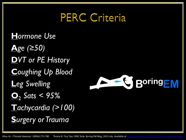
The Pulmonary Embolism Rule-Out Criteria was published in 2008 by Jeff Kline and is a second set of criteria to definitively rule-out PTE in patients ALREADY SCORE AS LOW RISK by Wells or Geneva.


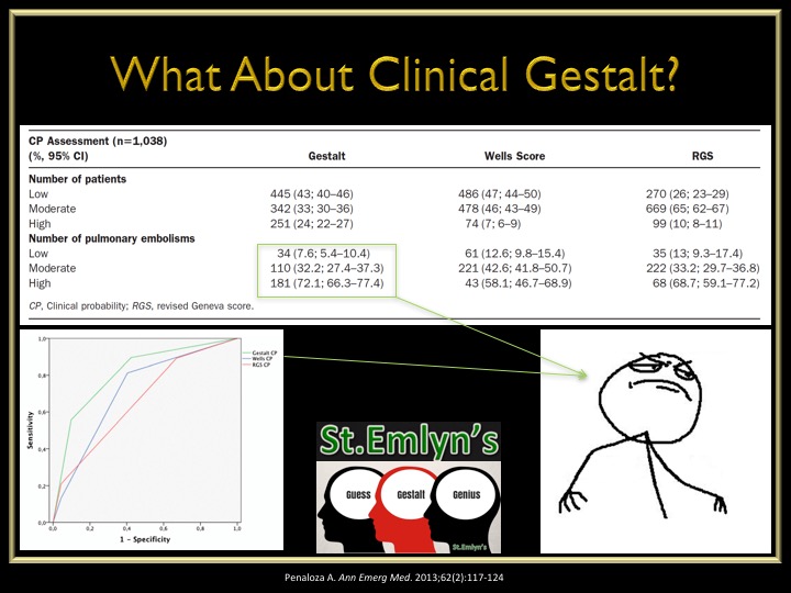
What about just good ol’ clinical gestalt?  An interesting study was performed in 2013 looking at the accuracy of Wells vs Geneva vs Gestalt and found:

  • Clinical gestalt had a lower missed rate of PTE in low-risk patients
  • Clinical gestalt had a high accuracy of diagnosing PTE in high-risk patients


The Work-Up of Suspected PTE

  1. Electrocardiogram is not senstitive nor specific for PTE but should be ordered on every patient with chest pain and/or shortness of breath to rule-out ACS
    1. The EMCMD talks about the 10 ECG findings of PTE in the best video I have every scene
  2. D-Dimer
    1. High senstivity = good for rule-out
    2. Should only be used after pre-test probability due to the false positives and unnecessary work-ups
    3. ADJUST-PE Study
      1. Found D-Dimer go up with age and created an age adjusted D-Dimer cutoff of:
        1. Age (yr) x 10 as diagnostic threshold
  3. Radiographic Imaging
    1. Computed tomography is gold standard but has higher radiation exposure and contrast loads
    2. Image result for CT PTE
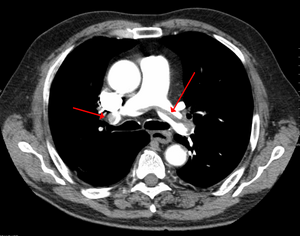
    3. Ventilation/Perfusion scan is safer in renal patients but up to 2/3rd are non-diagnostic
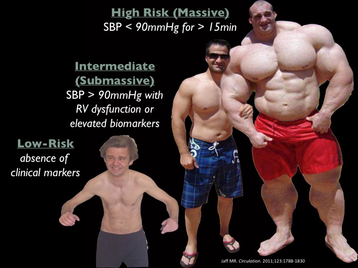
    4. Image result for V/Q PTE

Risk Assessment

Once you diagnose a patient with a PTE, you have determine the patient’s risk and severity of disease.

  1. Echocardiogram
    1. Looking for RV strain
      1. RV:LV ≥ 1
      2. RV hypokinesis
      3. Paradoxical septal movement
      4. Tricuspid regurgitation
  2. Biomarkers
    1. Brain Natriuretic Peptide (BNP)
      1. > 90 pg/mL has been associated with increased mortality
    2. Troponin
      1. > 0.01 ng/mL suggests evidence of RV dysfunction
  3. Pulmonary Embolism Severity Index (PESI)
    1. Published in 2005 and simplified 2010
    2. Developed to help prognosticate 30d mortality and found low-risk patients (PESI – 0) can be safely treated as outpatient


Definitions/Grades of PTE


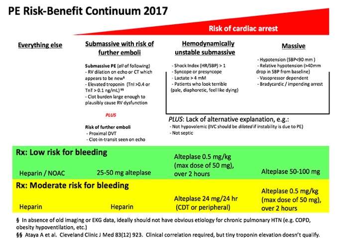
Treatment Strategies for PTE

  1. Anticoagulation
    1. Started with confirmation of PTE or with high pre-test probability during workup
    2. Lots of options (heparin, LMWH, direct thrombin inhibitors, Factor Xa inhibitors)
  2. Fibrinolytics
    1. Lots of recent research on who to lyse and who not to
    2. Original research showed benefit if full dose lytics were given to massive PTE, but harm in submassive patients
    3. This led to MOPPET in 2013 evaluating 1/2 dose lytics in submassive patients and found:
      1. Reduction in overall mortality
      2. No difference in bleeding complications
      3. Reduction in hospital stay
    4. PEITHO came next in 2014 and looked at full dose lytics vs anticoagulation only for submassive PTE and found:
      1. No mortality benefit
      2. Reduction in hemodynamic compromise
      3. Increase in major bleeding and intracranial hemorrhage
  3. Catheter Directed Therapy
    1. Good options in patients with a high risk of bleeding with systemic fibrinolytic therapy
    2. This can include:
      1. Focal fibrinolytic therapy at the clot
      2. Mechanical thombectomy
        1. AngioJet system
        2. AngioVac system
      3. Ultrasound Assisted Local Fibrinolytic Therapy
        1. ULTIMA and SEATTLE-II studies found reduction in RV:LV ratio and decreased bleeding complications
  4. Surgical Thrombectomy
    1. Can be used as a last resort option and mortality from these procedures has dramatically improved from 57% in the 1960s to < 6% in 2005

Putting It All Together

This is a graphic I modified from Jeff Kline and EmCrit that encompasses everything into a nice, neat package and as I have said before “algorithms will set you free”


References

  1. Beckman MG. Venous Thromboembolism: A Public Health Concern. Am J Prev Med. 2010;38(4S):S495-S501
  2. Spyropoulous AC. Direct medical costs of venous thromboembolism and subsequent hospital readmission rates: an administrative claims analysis from 30 managed care organizations. J Manag Care Pharm. 2007;13(6):475-486
  3. Calder KK. The mortality of untreated pulmonary embolism in emergency department patients. Ann Emerg Med. 2005;45(3):302-310
  4. Stein PD. Silent pulmonary embolism in patients with deep venous thrombosis: A systematic review. Am J Med. 2010;123:426-431
  5. Stein PD. Clinical Characteristics of patients with acute pulmonary embolism. Am J Med. 2007;120:871-879
  6. Pollack CV. Clinical characteristics, management, and outcoms of patients diagnosed with acute pulmonary embolism in the emergency department. JACC. 2011;57(6):700-706
  7. Wells PS. Use of a clinical model for safe management of patients with suspected pulmonary embolism. Ann Intern Med. 1998;129:997-1005
  8. Wells PS. Derivation of a simple clinical model to categorize patients probability of pulmonary embolism: increasing the models utility with the SimpliRED D-dimer. Thromb Haemost. 2000;83:416-420
  9. Wells PS. Excluding pulmonary embolism at the bedside without diagbnostic imaging: management of patients with suspected pulmonary embolism presenting to the emergency department by using a simple clinical model and D-dimer.  Ann Intern Med. 2001;135:98-107
  10. van Belle A. Effectiveness of managing suspected pulmonary embolism using an algorithm combining clinical probability, D-dimer testing, and computed tomography. JAMA. 2006;295(2):172-179
  11. Wicki J. Assessing clinical probability of pulmonary embolism in the emergency ward: a simple score. Arch Intern Med. 2001;161:997-92-97
  12. Le Gal G. Prediction of pulmonary embolism in the emergency department: the revised Geneve score. Ann Intern Med. 2006;144:165-171
  13. Klok FA. Simplication fo the revised Geneva score for assessing clinical probability of pulmonary embolism. Ann Intern Med. 2008;168(19):2131-2136
  14. Kline JA. Prospective multicenter evaluation of the pulmonary embolism rule-out criteria. J Thromb Haemost. 2008;6:772-780
  15. Penaloza A. Comparison of the unstructured clinical gestalt, the wells score, and the revised Geneva score to estimate pretest probability for suspected pulmonary embolism. Ann Emerg Med. 2013;62(2):117-124
  16. Righini M. Age-adjusted D-dimer cutoff levels to rule-out pulmonary embolism: the ADJUST-PE study. 2014;311(11):1117-1124
  17. Stein PD. Clinical characteristics of patients with acute pulmonary embolism: data from IOPED II. Am J Med. 2007;120:871-879
  18. Anderson DR. Computerized tomographic pulmonary angiography versus ventilation perfusion lung scanning for the diagnosis of pulmonary embolism. Curr Opin Pulm Med. 2009;15:425–429
  19. Rudoni RR. Use of two-dimensional echocardiography for the diagnosi of pulmonary embolus. J Emerg Med. 1998;16(1):5-8
  20. Taylor RA. Point-of-care focused cardiac ultrasound for prediction of pulmonary embolism adverse outcomes. J Emerg Med. 2013;45(3):392-399
  21. Kiely DG. Elevated levels of natriuretic peptides in patients with pulmonary thromboembolism. Resp Med. 2005;99:1286-1291
  22. Jaff MR. Management of massive and submassive pulmonary embolism, iliofemoral deep vein thrombosis, chronic thromboembolic pulmonary hypertension: a scientific statement form the American Heart Association. Circulation. 2011;123:1788-1830
  23. Keller K. Cardiac troponin I for predicting right ventricular dysfunction and intermediate risk in patients with normotensive pulmonary embolism. Neth Heart J. 2015;23:55-61
  24. Aujesky D. Derivation and validation of a prognostic model for pulmonary embolism. Am J Respir Crit Care Med. 2005;172:1041-1046
  25. Jimenez D. Simplification of the pulmonary embolism severity index for prognostication in patients with acute symptomatic pulmonary embolism. Arch Intern Med. 2010;170(15):1383-1389
  26. Tapson VF. Treatment of pulmonary embolism: anticoagnulation, thrmbolytic therapy, and complications of therapy. Crit Care Clin. 2011;27:825-839
  27. Sharifi M. Moderate pulmonary embolism treated with thrombolysis. Am J Cardiol. 2013;111:273-277
  28. Zhang Z. Lower dosage of recombinant tissue-type plasminogen activator (rt-PA) in the treatment of acute pulmonary embolism: a systematic review and meta-analysis. Thrombosis Research. 2014;133:357-363
  29. Meyer GM. Fibrinolysis for patients with intermediate-risk pulmonary embolism. NEJM. 2014;370(15):1402-1411
  30. Chatterjee S. Thrombolysis for pulmonary embolism and risk of all-cause mortality, major bleeding, and intracranial hemorrhage: a meta-analysis. JAMA. 2014;311(23):2414-2421
  31. Curtis GM. Risk factors associated with bleeding after alteplase administration for pulmonary embolism: a case control study. 2014;34(8):818–825
  32. Kennedy RJ. Thrombus resolution and hemodynamic recovery using ultrasound-accelerated thrombolysis in acute pulmonary embolism. J  Vasc Interv  Radiol. 2013;24:841-848
  33. Kucher N. Randomized, controlled trial of ultrasound-assisted catheter-directed thrombolysis for acute intermediate-risk pulmonary pulmonary embolism. Circulation. 2014;129:479-486
  34. Cross FS. A survey of the current status of pulmonary embolectomy for massive pulmonary embolism. Circulation. 1967;35:186-191
  35. Stulz P. Decision making in the surgical treatment of massive pulmonary embolism. Eur J Cardio-thorac Surg. 1994;8:188-193
  36. Leacche M. Modern surgical treatment of massive pulmonary embolism: result in 47 sonsecutive patients after rapid diagnosis and aggressive surgical approach. J Thorac Cardiovasc Surg. 2005;129:1018-1023
  37. Prandoni P, et al (PESIT Investigators). Prevalance of Pulmonary Embolism amount Patients Hospitalized for Syncope.  NEJM.  2016;375:1524-1531
  38. John MA, Klok FA, van Es N. D-dimer Interval Likelihood Ratios for Pulmonary Embolism.  Acad Emerg Med.  2017;4;1-5.
  39. Konstantinides SV, et al. Impact of Thrombolytic Therapy on the Long-Term Outcome of Intermediate-Risk Pulmonary Embolism.  JACC.  2017;69(12):1536-1544
  40. Sharifi M, et al (PEAPETT Investigators). Pulseless electrical activity in pulmonary embolism treated with thrombolysis.  Am J Emerg Med.  2016;34(10):1963-1967
  41. Piazza G, et al. (SEATTLE-II Investigators). A prospective, single arm, multicenter trial of ultrasound-facilitated , catheter-directed, low-dose fibrinolysis for acute massive and submassive pulmonary embolism.  JACC. 2015;8(1):1382-1392

Ep-PAINE-nym


Kussmaul Breathing


Other Known AliasesNone

DefinitionForm of hyperpnea (hyperventilation) characterized by a rhythmic, labored, and deep respiration pattern


Clinical Significance Compensatory mechanism of profound metabolic acidosis, classically associated with diabetic ketoacidosis

History – Named after Adolph Kussmaul (1822-1902), a German physician, who noticed it in patients with severe diabetes mellitus and first published the finding in 1874.  Dr. Kussmaul was a prolific physician in the late 1880’s and this is just one of many eponymous distinctions that bears his name.  I am sure his name will come again in this series.


References

  1. Firkin BG and Whitwirth JA.  Dictionary of Medical Eponyms. 2nd ed.  New York, NY; Parthenon Publishing Group. 1996.
  2. Bartolucci S, Forbis P.  Stedman’s Medical Eponyms.  2nd ed.  Baltimore, MD; LWW.  2005.
  3. Yee AJ, Pfiffner P. (2012).  Medical Eponyms (Version 1.4.2) [Mobile Application Software].  Retrieved http://itunes.apple.com.
  4. Whonamedit – dictionary of medical eponyms. http://www.whonamedit.com/
  5. A. Kussmaul: Zur Lehre vom Diabetes mellitus. Über eine eigenthümliche Todesart bei Diabetischen, über Acetonämie, Glycerin-Behandlung des Diabetes und Einspritzungen von Diastase in’s Blut bei dieser Krankheit., Deutsches Archiv für klinische Medicin, Leipzig, 1874, 14: 1-46.
  6. Young P, Finn BC, Bruetman JE, Buzzi A, Zylberman M. [The outstanding achievements of Adolf Kussmaul]. Revista medica de Chile. 2012; 140(4):538-44. [pubmed]

Ep-PAINE-nym


Varess Needle


Other Known AliasesNone

Definition12-15cm long, 2 cannula instrument used for insuflating the abdominal cavity before laparoscopic port placement.  The outer cannula has a beveled needle for dissecting through the abdominal wall.  The spring-loaded inner stylet resides within the outer cannula and has a dull tip to prevent injury to abdominal viscera.  Due to this spring-loaded mechanism, the inner stylet retracts into the outer cannula while it moves through the abdominal planes and advances past the sharp, cutting tip of the outer cannula once through the peritoneum.

Image result for veress needleImage result for veress needle

Clinical Significance Using the Varess needle is the oldest and most traditional techniques for obtaining laparoscopic access

History – Named after János Vares (1903-1979), a Hungarian internist, who used iatrogenic pneumothoraces to treat tuberculosis patients.  He created this spring loaded needle in 1932 and published his results in 1936 (in a Hungarian journal), which was subsequently translated and published in German for wider audience in 1938.  Raoul Palmer (1904-1985), a French gynecologist, began using the Varess needle for laparoscopic surgery in 1947.


References

  1. Firkin BG and Whitwirth JA.  Dictionary of Medical Eponyms. 2nd ed.  New York, NY; Parthenon Publishing Group. 1996.
  2. Bartolucci S, Forbis P.  Stedman’s Medical Eponyms.  2nd ed.  Baltimore, MD; LWW.  2005.
  3. Yee AJ, Pfiffner P. (2012).  Medical Eponyms (Version 1.4.2) [Mobile Application Software].  Retrieved http://itunes.apple.com.
  4. Whonamedit – dictionary of medical eponyms. http://www.whonamedit.com/
  5. Vares J. Neues instrument zur ausfuhrung von brust-oder bauchpunktionen und pneumothoraxbehandlung. Deut Med Wochenschr. 1938;64:1480-1481.
  6. Palmer R. Instrumentation et technique de la coelioscopie gynécologique. Gynecologie et obstetrique. 1947; 46(4):420-31. [pubmed]

PAINE #PANCE Pearl – Surgery


Question


  1. What are the five classic causes of a post-operative fever?
  2. What are two other clever causes to think of (if I do say so myself)?


Answer


  1. The five classic causes of post-operative fever are:
    1. Wind = lungs (atelectasis, pneumonia, aspiration) = POD 1-2
    2. Water = UTI = POD 2-3
    3. Wound = surgical site infection = POD 3-5
    4. Walking = DVT = POD 3-5
    5. Wonder drugs = drug reactions = anytime
  2. Another two “W’s” to add to this list:
    1. Withdrawal = typically alcohol
    2. “Wonky” glands = thyrotoxicosis, adrenal crisis


Check out my article in JAAPA from 2016 on “Evaluating Postoperative Fever” for a more in-depth look

Ep-PAINE-nym


Glisson’s Capsule


Other Known AliasesNone

DefinitionOuter capsule of connective fibrous tissue, surrounding the liver, the intrahepatic branches of the portal vein, hepatic arteries, and bile duct

Clinical Significance The is a structure that must be dissected while operating on the liver.  In trauma, you can have subcapsular hematomas from hemorrhage that are contained by Glisson’s capsule.

History – Named after Francis Glisson (1597-1667), who was an English physician, anatomist, and pathologist.  His work on the liver in the late 1600s produced the foremost textbook on the digestive system, The Anatomia Hepatis, where he first described the covering of the liver in detail.


References

  1. Firkin BG and Whitwirth JA.  Dictionary of Medical Eponyms. 2nd ed.  New York, NY; Parthenon Publishing Group. 1996.
  2. Bartolucci S, Forbis P.  Stedman’s Medical Eponyms.  2nd ed.  Baltimore, MD; LWW.  2005.
  3. Yee AJ, Pfiffner P. (2012).  Medical Eponyms (Version 1.4.2) [Mobile Application Software].  Retrieved http://itunes.apple.com.
  4. Whonamedit – dictionary of medical eponyms. http://www.whonamedit.com/
  5. Haubrich WS. Glisson of Glisson’s capsule of the liver. Gastroenterology. 2001; 120(6):1362. [pubmed]

#31 – Small Bowel Obstruction


***LISTEN TO THE PODCAST HERE***


Epidemiology

Small bowel obstruction (SBO) is one of the more common surgical emergencies in US, due to the increased intraluminal pressures which can lead to intestinal ischemia and risk of spontaneous rupture.  Overall mortality increases significantly if either of these occur.  It is estimated that over 300,000 surgeries per year occur in the US for bowel obstructions, with the small bowel making up around 80%.  Although it can happen at age, there is a higher trend to older patients and thee is equal incidence with both genders.


Risk Factors

The most common cause of SBO is postoperative adhesions, but there are numerous other causes that you should keep on your differential.


Signs and Symptoms

  • Abdominal pain
    • Paroxysms of periumbilical pain (3-5 minutes)
  • Nausea
  • Vomiting
  • Obstipation

Physical Examination

  • Dehydration
    • Tachycardia, orthostasis, decreased urine output
  • +/- Distension
  • Changes in bowel sounds
    • Hyperactive à muffled à absent
  • Percussion changes
    • Tympanic with distension
    • Dullness with fluid
  • Peritoneal signs if perforation present
  • +/- hemoccult

Laboratory Studies

  • CBC with differential
    • Anemia can point to a chronic condition
  • BMP
  • ABG
    • Acidosis = bowel ischemia, volume depletion
    • Alkalosis = vomiting
  • Serum lactate

Imaging Studies

  • Plain Radiographs
    • Dilated loops of small bowel with air/fluid levels on upright film
    • May also see a paucity of gas in distal intestines
    • Small bowel dilation ≥ 2.5cm is diagnostic

  • Abdominal CT
    • Much more sensitive and can also identify the specific causes
    • Can also identify a transition point
    • Other radiological signs on CT consistent with SBO include:
      • Bowel wall thickening > 3mm
      • Submucosal edema
      • Mesenteric edema
      • Ascites
      • Target sign (intussusception)
      • Whirl sign (volvulus)
      • Venous cut-off sign (thrombosis)
  • Ultrasound
    • Not as good as CT, but better than plain radiography
  • Special Considerations on Radiography
    • Closed-Loop Obstruction
      • High risk for ischemia, perforation, and ischemic bowel
    • Ischemia and Perforation

Management

  • Initial
    • NPO
    • Fluid resuscitation
    • Surgery consultation
    • Gastrointestinal decompression
      • Not in EVERY patient, but good for patients with significant distension, nausea, and/or vomiting
    • Surgery
      • Nonspecific signs of bowel ischemia:
        • Fever
        • Leukocytosis
        • Tachycardia
        • Continuous and/or worsening abdominal pain
        • Metabolic acidosis
        • Peritonitis
      • High likelihood of bowel resection if ≥ 3 of the following:
        • Pain > 4 days
        • Abdominal guarding on exam
        • Elevated CRP > 75 mg/dL
        • Leukocytosis > 10,000
        • > 500cc fluid of intraabdominal fluid
        • Reduced wall contrast enhancement on CT
    • Observation
      • If no high-risk surgical signs present, observation with serial examinations may be used for 12-24 hours in patients with:
        • Early postoperative obstruction
        • Inflammatory bowel disease
        • Gallstone ileus
        • Infectious small bowel disease
        • Colonic diverticular disease
      • Exploration should proceed if no improvement

References

  • Miller G, Boman J, Shrier I, Gordon PH. Etiology of small bowel obstruction. American Journal of Surgery. 2000; 180(1):33-6. [pubmed]
  • Markogiannakis H, Messaris E, Dardamanis D. Acute mechanical bowel obstruction: clinical presentation, etiology, management and outcome. World Journal of Gastroenterology. 2007; 13(3):432-7. [pubmed]
  • Scott FI, Osterman MT, Mahmoud NN, Lewis JD. Secular trends in small-bowel obstruction and adhesiolysis in the United States: 1988-2007. American Journal of Surgery. 2012; 204(3):315-20. [pubmed]
  • Drożdż W, Budzyński P. Change in mechanical bowel obstruction demographic and etiological patterns during the past century: observations from one health care institution. Archives of Surgery (Chicago, Ill. : 1960). 2012; 147(2):175-80. [pubmed]
  • Taylor MR, Lalani N. Adult small bowel obstruction. Academic Emergency Medicine. 2013; 20(6):528-44. [pubmed]
  • Jackson PG, Raiji MT. Evaluation and management of intestinal obstruction. American Family Physician. 2011; 83(2):159-65. [pubmed]
  • Mullan CP, Siewert B, Eisenberg RL. Small bowel obstruction. American Journal of Roentgenology. 2012; 198(2):W105-17. [pubmed]
  • Suri S, Gupta S, Sudhakar PJ, Venkataramu NK, Sood B, Wig JD. Comparative evaluation of plain films, ultrasound and CT in the diagnosis of intestinal obstruction. Acta radiologica. 1999; 40(4):422-8. [pubmed]
  • Zalcman M, Sy M, Donckier V, Closset J, Gansbeke DV. Helical CT signs in the diagnosis of intestinal ischemia in small-bowel obstruction. American Journal of Roentgenology. 2000; 175(6):1601-7. [pubmed]
  • Eltarawy IG, Etman YM, Zenati M, Simmons RL, Rosengart MR. Acute mesenteric ischemia: the importance of early surgical consultation. The American Surgeon. 2009; 75(3):212-9. [pubmed]
  • Schwenter F, Poletti PA, Platon A, Perneger T, Morel P, Gervaz P. Clinicoradiological score for predicting the risk of strangulated small bowel obstruction. The British Journal of Surgery. 2010; 97(7):1119-25. [pubmed]

PAINE #PANCE Pearl – Surgery Edition


Question


What are the boundaries of Calot’s Triangle and what can be found there?


Answer


Calot’s Triangle is the area bordered by:

  1. Cystic duct
  2. Common hepatic duct
  3. Cystic artery

A lymph node can be found within this triangle and there is apparently great debate about the name of this node.   I had learned this was referred to as Calot’s node (which makes sense, right), but it is more accurately named Lund’s, or Mascagni’s, node.  This node can be enlarged and inflamed with cholecystitis.

The anatomical area is referred to as the cystohepatic triangle and is bordered by:

  1. Cystic duct
  2. Common hepatic artery
  3. Superior, inferior margin of the liver

The reason for this distinction from Calot’s Triangle is because the cystic artery can be found within this region.

Blue = Calot’s and Red = Cystohepatic


References

  1. Blackbourne LH.  Surgical Recall.  6th Edition.  2012.
  2. Haubrich WS. Calot of the triangle of Calot. Gastroenterology. 2002; 123(5):1440. [pubmed]
  3. Miranda, Efrain A., PhD. “Triangle of Calot.” Medical Terminology Daily. N.p., 10 May 2016. Web. 10 June 2017.