Ep-PAINE-nym


Apt-Downey Test


Other Known Aliases – alkali denaturation test

DefinitionLab test where the blood sample is hemolyzed using sterile water and centrifuged to produce a hemoglobin supernatent.  This is then mixed with 1% NaOH.  Fetal hemoglobin will stay pink, while maternal hemoglobin will turn yellow/brown.

fig-2-the-complete-vasa-previa-testing-setup-shown-a-beaker-of-014-m-naoh-and-two

Clinical SignificanceHelps differentiate maternal from fetal blood in cases of vaginal bleeding or neonatal hematemesis/hematochezia.

History – Given to Dr. Leonard Apt and Dr. William Downey, Jr., who were physicians at Harvard Medical School and Children’s Medical Center in Boston, MA in the mid-1900’s.


References

  1. Firkin BG and Whitwirth JA.  Dictionary of Medical Eponyms. 2nd ed.  New York, NY; Parthenon Publishing Group. 1996.
  2. Yee AJ, Pfiffner P. (2012).  Medical Eponyms (Version 1.4.2) [Mobile Application Software].  Retrieved http://itunes.apple.com
  3. Apt L, Downey WS. Melena neonatorum: the swallowed blood syndrome; a simple test for the differentiation of adult and fetal hemoglobin in bloody stools. The Journal of Pediatrics. 47(1):6-12. 1955. [pubmed]

PAINE #PANCE Pearl – OB/GYN


  1. Women should be screened for gestational diabetes at 24-28 gestation.
  2. There are 2 different recommended testing strategies:
    1. Two-Step (most common)
      1. 50g glucose challenge
        1. Given regardless of when last meal was.
        2. Serum glucose measured at 1-hour
          1. ≥130-140 mg/dL is positive test
            1. The lower the threshold the higher sensitivity, but increased false positives
            2. The higher the threshold the higher the specificity, but with decreased sensitivity
      2. If 1st step positive, a 100g glucose challenge given
        1. Overnight fast and measured at:
          1. Fasting –> (+) if ≥ 95 mg/dL
          2. 1 hour –> (+) if ≥ 180 mg/dL
          3. 2 hour –> (+) if ≥ 155 mg/dL
          4. 3 hour –> (+) if ≥ 140 mg/dL
    2. One-Step
      1. 75 glucose challenge
        1. Given after overnight fast and measured at:
          1. Fasting –> (+) if ≥ 92 mg/dL
          2. 1 hour –> (+) if ≥ 180 mg/dL
          3. 2 hour –> (+) if ≥ 153 mg/dL

References

  1. Practice Bulletin No. 137: Gestational diabetes mellitus. Obstetrics and Gynecology. 2013;122(2 Pt 1):406-16. [pubmed]
  2. Hod M, Kapur A, Sacks DA. The International Federation of Gynecology and Obstetrics (FIGO) Initiative on gestational diabetes mellitus: A pragmatic guide for diagnosis, management, and care. International journal of gynaecology and obstetrics. 2015;131 Suppl 3:S173-211. [pubmed]
  3. Moyer VA, . Screening for gestational diabetes mellitus: U.S. Preventive Services Task Force recommendation statement. Annals of internal medicine. 204;160(6):414-20. [pubmed]

Ep-PAINE-nym


Ferguson’s Reflex


Other Known AliasesFetus Ejection Reflex

DefinitionStimulation of the cervix leading to contraction of the uterus through oxytocin release.

Clinical SignificanceDemonstrates positive feedback during labor and delivery to increase uterine contractions as cervical dilation progresses. Long standing belief that epidural anesthesia before cervical dilation increased risk of cesarean section (this was debunked in 2005).

History – Given to Dr. James Ferguson, a Canadian obstetrician, in 1940 after he showed this physiologic effect in rabbits and postulated it to be true in humans.


References

  1. Firkin BG and Whitwirth JA.  Dictionary of Medical Eponyms. 2nd ed.  New York, NY; Parthenon Publishing Group. 1996.
  2. Wong CA, Scavone BM, Peaceman AM. The risk of cesarean delivery with neuraxial analgesia given early versus late in labor. The New England journal of medicine. 2005;352(7):655-65. [pubmed]
  3. Newton N.  The fetus ejection reflex revisited.  Birth.  1987;14(2):106-108.

#27 – Abnormal Uterine Bleeding – Definitions and Classifications


***LISTEN TO THE PODCAST HERE***


History of FIGO

Because of confusing terminology and difficulty in translating to other languages, the International Federation of Obstetrics and Gynecology (FIGO) created a special task force in 2005 charged with clarifying the terminology and classifying the different causes.  This way clinicians, patients, and researchers throughout the world could be talking the same language.  Dysfunctional uterine bleeding (DUB) was replaced by abnormal uterine bleeding (AUB).. They also introduced a classification system to help sub-divide the causes of AUB.


But in order to define something as “abnormal”, they needed to define normality, which can obviously be very difficult when incorporating the world’s population. The consensus definitions were then agreed upon in 2015 to include the 5th to 95th percentiles form the available data.


The FIGO definition of AUB is any symptomatic variation from normal menstruation, with regards to frequency, regularity, duration, or volume.

screen-shot-2017-02-02-at-4-21-49-pm


Classifications of Abnormal Uterine Bleeding

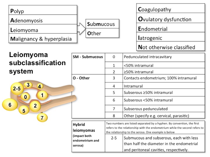
In 2011, FIGO created a classification system for the main causes of AUB.  It is broken down into 2 main categories based on whether or not the pathology can be seen on imaging or histopathology.  This also allows for subclassifications due to multiple etiologies.

  • Structural (PALM)
    • Polyps (AUB-P)
    • Adenomyosis (AUB-A)
    • Leiomyomas (AUB-L)
      • Hierarchy of classification
        • Primary
          • Presence or absence
        • Secondary
          • Submucosal
            • Abuts the endometrium or distorts the endometrial cavity
          • Other
            • Subserosal
          • Tertiary
            • 0-8 numbering system based on endometrial or serosal involvement
            • Hybrid (2-5)
              • Submucosal and subserosal
    • Malignancy and hyperplasia (AUB-M)
  • Non-structural (COEIN)
    • Coagulopathy (AUB-C)
      • Most commonly is von Willebrand disease
    • Ovulatory dysfunction (AUB-O)
      • At least one cycle that varies by more than 7 days in 12 months
    • Endometrial (AUB-E)
      • Category of exclusion
    • Iatrogenic (AUB-I)
      • Medications
        • Anticoagulants
        • Hormone therapies
      • IUDs
    • Not otherwise classified (AUB-N)

picture1


Documentation

Very similar to the documentation for an OB patient (TPAL score), the documentation uses the PALM-COEIN scoring system for “simplicity”.  Example:

  • Patient with adenomyosis would be:
    • P0A1L0M0-C0O0E0I0N0
  • Patient with endometrial hyperplasia and a subserosal leiomyoma < 50% intramural would be:
    • P0A0L6M1-C0O0E1I0N0

References

  1. Fraser IS, Critchley HO, Munro MG, Broder M, . A process designed to lead to international agreement on terminologies and definitions used to describe abnormalities of menstrual bleeding. Fertility and Sterility. 2007; 87(3):466-76. [pubmed]
  2. Woolcock JG, Critchley HO, Munro MG, Broder MS, Fraser IS. Review of the confusion in current and historical terminology and definitions for disturbances of menstrual bleeding. Fertility and Sterility. 2008;90(6):2269-80. [pubmed]
  3. Fraser IS, Critchley HO, Munro MG, Broder M. Can we achieve international agreement on terminologies and definitions used to describe abnormalities of menstrual bleeding? Human reproduction (Oxford, England). 2007;22(3):635-43. [pubmed]
  4. Harlow SD, Lin X, Ho MJ. Analysis of menstrual diary data across the reproductive life span applicability of the bipartite model approach and the importance of within-woman variance. Journal of clinical epidemiology. 2000;53(7):722-33. [pubmed]
  5. Fraser IS, Critchley HO, Broder M, Munro MG. The FIGO recommendations on terminologies and definitions for normal and abnormal uterine bleeding. Seminars in reproductive medicine. 2011;29(5):383-90. [pubmed]
  6. Munro MG, Critchley HO, Broder MS, Fraser IS, . FIGO classification system (PALM-COEIN) for causes of abnormal uterine bleeding in nongravid women of reproductive age. International journal of gynaecology and obstetrics: the official organ of the International Federation of Gynaecology and Obstetrics. 2011;113(1):3-13. [pubmed]

Ep-PAINE-nym


Chadwick Sign


DefinitionBlue discoloration of the cervix and vaginal mucosa seen in early pregnancy.

Clinical SignificanceDue to increased blood flow to support a newly implanted embryo and can be seen 6-8 weeks after conception.

History – Given to James R. Chadwick, an American gynecologist of the late 19th century, after he read a paper before the American Gynecologic Society in 1886, but 1st described by Etienne Jacquemin in 1836.  Dr. Chadwick did give appropriate recognition to Dr. Jacquemin during this presentation.


References

  1. Firkin BG and Whitwirth JA.  Dictionary of Medical Eponyms. 2nd ed.  New York, NY; Parthenon Publishing Group. 1996.
  2. Gleichert JE. Etienne Joseph Jacquemin, discoverer of ‘Chadwick’s sign’. Journal of the history of medicine and allied sciences. 1971;26(1):75-80. [pubmed]

PAINE #PANCE Pearl – Cardiovascular


82-year-old male, with a history of HTN, HLD, and CAD, presents to your clinic with a six-month history of dyspnea on exertion.  He states he is unable to walk as far as he used when exercising, and when he over exerts himself, he reports having some mild chest pain and feeling lightheaded.  This resolves with rest and he denies any syncope with these events.


Medications

Metoprolol 50mg daily

Lisinopril 10mg daily

Simvastatin 30mg daily


Vital Signs

BP – 158/97

HR – 62

RR – 13

O2% – 100%


Physical exam

General – WN/WD male in NAD

Pulmonary – CTA without adventitial breath sounds

CV – Soft S2 with murmur over right 2nd intercostal space

PV – carotid pulse is weak and has a slow rise, murmur is appreciated

Neuro – No focal deficits


EKG

lvh


This patient has aortic stenosis.  The suggestive parts of the H&P are:

  • History
    • The classic triad of aortic stenosis is chest pain, dyspnea, and syncope.
  • Aortic stenosis increases in prevalence with age
  • Cardiac Auscultation
    • Soft, single S2 since A2, which is due to aortic valve closure, is delayed and occurs with P2
    • Murmur
      • Systolic ejection murmur best heard over the right 2nd intercostal space
      • medicosnotes_heart-sounds-and-murmur-in-aortic-stenosis
      • Begins on S1 and ends before S2
      • May radiate to the carotids
      • 634464_xlarge
  • Peripheral Vascular
    • Carotid Palpation
    • Pulsus Parvus et Tardus (weak and late)
    • pulse-jvp-12-638
  • EKG
    • Shows LVH and strain pattern in precordial leads

  1. What is the next step in the management of this patient?
    1. Transthoracic echocardiography
  2. After the next step, what important variables must you specifically assess?
    1. Valvular anatomy and size
      1. Aortic valve surface area
    2. Valve hemodynamics
      1. Transvavular aortic velocity
      2. Mean transvalvular pressure
    3. LV size and ejection fraction
    4. Pulmonary artery pressure
    5. Other concomitant conditions


These variables will help with staging the severity of the stenosis, as well as determine need for operative intervention.

Ep-PAINE-nym


Bainbridge Reflex


Other known aliasesAtrial Reflex

DefinitionCompensatory increase in heart rate caused by a rise in right atrial pressure.  Opposite of carotid baroreceptors.

Clinical SignificanceRespiratory sinus arrythmia.  Inspiration causes increased venous return.

History – Described by Francis Arthur Bainbridge in 1915


References

  1. Hakumäki MO. Seventy years of the Bainbridge reflex. Acta physiologica Scandinavica. 1987;130(2):177-85. [pubmed]
  2. Firkin BG and Whitwirth JA.  Dictionary of Medical Eponyms. 2nd ed.  New York, NY; Parthenon Publishing Group. 1996.
  3. http://www.healio.com/cardiology/learn-the-heart/cardiology-review/topic-reviews/bainbridge-reflex

PAINE #PANCE Pearl – Cardiovascular

82-year-old male, with a history of HTN, HLD, and CAD, presents to your clinic with a six-month history of dyspnea on exertion.  He states he is unable to walk as far as he used when exercising, and when he over exerts himself, he reports having some mild chest pain and feeling lightheaded.  This resolves with rest and he denies any syncope with these events.


Medications

Metoprolol 50mg daily

Lisinopril 10mg daily

Simvastatin 30mg daily


Vital Signs

BP – 158/97

HR – 62

RR – 13

O2% – 100%


Physical exam

General – WN/WD male in NAD

Pulmonary – CTA without adventitial breath sounds

CV – Soft S2 with murmur over right 2nd intercostal space

PV – carotid pulse is weak and has a slow rise, murmur is appreciated

Neuro – No focal deficits


EKG

lvh


Questions

  1. What is the next step in the management of this patient?
  2. After the next step, what important variables must you specifically assess?

#26 – Pericardial Effusion and Cardiac Tamponade


***LISTEN TO THE PODCAST HERE***


Anatomy

The pericardium consists of a double-layered semi-elastic sac that holds the heart in the mediastinum.  Basically, so the heart doesn’t flop around inside the thoracic cavity.  There should be a small amount of fluid (15-50mL) present to prevent adhesion of the pericardial sac to the heart.  It is then termed an effusion when it is more than the normal amount.  How much quantifies an effusion?  Doesn’t matter…. what does matter is how fast that fluid develops.  Because the pericardium is semi-elastic, it can accommodate and stretch over time if the accumulation is slow.  This would lead to a greater volume of fluid before symptoms occur.  If the fluid accumulates rapidly, less volume can produce profound effects due to the restrictive nature of the fibrous pericardium.

picture1

picture1


Etiology

  • Infectious
    • Viral
    • Bacterial
    • Fungal
    • Parasitic
  • Non-infectious
    • Neoplastic
    • Autoimmune/inflammatory
    • Trauma
    • Cardiac
    • Radiation
    • Metabolic

Signs and Symptoms

There are no reliable historical clues or physical exam findings that are specific to pericardial effusions.  They are helpful, though, to sort out the cause of the effusion. Common findings include:

  • Fever
  • Dyspnea
  • Chest pain
  • Tachycardia
  • JVD
  • Hepatomegaly
  • Pulsus paradoxus
  • Ewart’s Sign
    • Dullness to percussion, egophony, and bronchial breath sounds over the inferior angle of the left scapula
  • Beck’s Triad
    • Hypotension
    • JVD
    • Muffled heart tones

Work-Up

  • EKG
  • Chest X-ray
    • Small effusions are generally not appreciated on radiography
    • Larger, chronic effusions may appear as an enlarged cardiac silhouette classically referred to as a “Water bottle heart”
    • pericardial-effusion-water-bottle-sign-1

      Radiopaedia

  • Echocardiogram
    • Looking for anechoic stripe around the heart
    • 2D Apical 4-chamber is my view of choice
    • Severity can also be assessed by looking for:
      • RV collapse during diastole
      • LV collapse with increased EF
      • IVC dilation and loss of respiratory variations
    •  

ccen1iwusaevl0l-jpg_large


Treatment

  • Pericardiocentesis with catheter placement
    •  

    • Three-way stopcock is used to measure pericardial pressure
    • Fluid is then sequentially removed and pressure re-measured until < 5mmHg during inspiration
    •  

  • Open surgical drainage via pericardial window if:
    • Fluid accumulates after catheter drainage
    • Effusion is loculated
    • Need for biopsy
    • Patient has coagulopathy
    •  

    •  


References

  1. Braunwald E. Pericardial Disease. In: Kasper D, Fauci A, Hauser S, Longo D, Jameson J, Loscalzo J. eds. Harrison’s Principles of Internal Medicine, 19e. New York, NY: McGraw-Hill; 2015. http://accessmedicine.mhmedical.com/content.aspx?bookid=1130&Sectionid=79743215. Accessed January 12, 2017.
  2. Imazio M. Contemporary management of pericardial diseases. Current Opinion in Cardiology. 2012;27(3):308-17. [pubmed]
  3. Levy PY, Corey R, Berger P. Etiologic diagnosis of 204 pericardial effusions. Medicine. 2003;82(6):385-91. [pubmed]
  4. Permanyer-Miralda G. Acute pericardial disease: approach to the aetiologic diagnosis. Heart (British Cardiac Society). 2004;90(3):252-4. [pubmed]
  5. Bruch C, Schmermund A, Dagres N. Changes in QRS voltage in cardiac tamponade and pericardial effusion: reversibility after pericardiocentesis and after anti-inflammatory drug treatment. Journal of the American College of Cardiology. 2001;38(1):219-26. [pubmed]
  6. Sternbach G. Claude Beck: cardiac compression triads. The Journal of Emergency Medicine. 1989;6(5):417-9. [pubmed]
  7. Stanford University. Tamponade. Echocardiography in ICU. https://web.stanford.edu/group/ccm_echocardio/cgi-bin/mediawiki/index.php/Tamponade.
  8. Adler Y, Charron P, Imazio M. 2015 ESC Guidelines for the diagnosis and management of pericardial diseases: The Task Force for the Diagnosis and Management of Pericardial Diseases of the European Society of Cardiology (ESC)Endorsed by: The European Association for Cardio-Thoracic Surgery (EACTS). European Heart Journal. 2015;36(42):2921-64. [pubmed]
  9. Gumrukcuoglu HA, Odabasi D, Akdag S, Ekim H. Management of Cardiac Tamponade: A Comperative Study between Echo-Guided Pericardiocentesis and Surgery-A Report of 100 Patients. Cardiology Research and Practice. 2011:197838. [pubmed]