Ep-PAINE-nym


Cheyne-Stokes Respirations

Other Known Aliasesnone

Definitionoscillating, crescendo-decrescendo pattern of progressive deeper and faster breathing followed a gradual decrease culminating in a period of apnea

Clinical Significance this pattern is theorized to be a delay in changes to ventilation after detection of PaCO2 changes. This lag causes the classic respiratory pattern. Conditions associated with this include cardiac disease, neurologic disease, sedation, acid-base disturbances, prematurity in infancy, and rapid altitude changes.
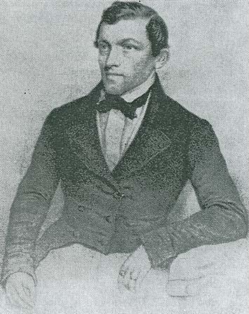
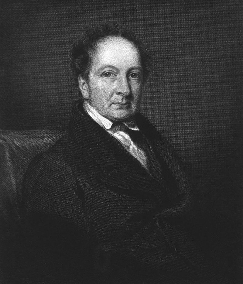
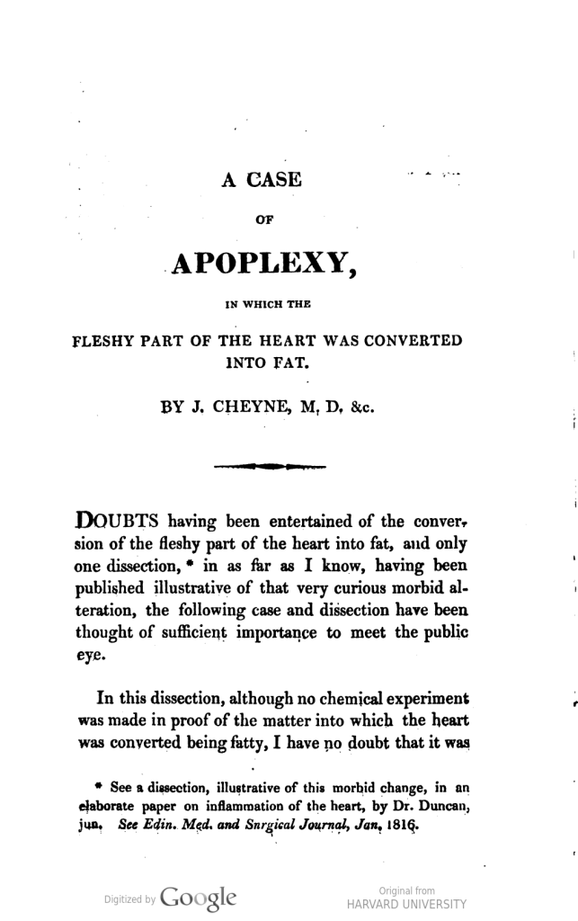
HistoryNamed after John Cheyne (1777-1836) , who was a British surgeon and received his medical doctorate at the age of 18 from Edinburgh University. He would serve as a military surgeon for several years before joining his father’s medical practice and ultimately, moving to Dublin for the majority of his career. Some have credited him as “The Father of Medicine in Ireland”. He would describe his eponymous findings in his 1818 article entitled ” A case of apoplexy in which the fleshy part of the heart was converted to fat”

William Stokes (1804-1878), was an Irish physician and received his medical doctorate from the University of Edinbugh in 1825. He was a leader and pioneer in the adaptation of the Parisian school of anatomical diagnosis and helped introduce the stethoscope to clinical practice in Ireland. He would note his eponymous findings in his 1854 textbook entitled ” The Diseases of the Heart and Aorta” and cited Dr. Cheyne as observing this first.


References

  1. Firkin BG and Whitwirth JA.  Dictionary of Medical Eponyms. 2nd ed.  New York, NY; Parthenon Publishing Group. 1996.
  2. Bartolucci S, Forbis P.  Stedman’s Medical Eponyms.  2nd ed.  Baltimore, MD; LWW.  2005.
  3. Yee AJ, Pfiffner P. (2012).  Medical Eponyms (Version 1.4.2) [Mobile Application Software].  Retrieved http://itunes.apple.com.
  4. Whonamedit – dictionary of medical eponyms. http://www.whonamedit.com
  5. Up To Date. www.uptodate.com
  6. Cheyne J. A case of apoplexy in which the fleshy part of the hear was converted into fat. Dubin Hospital Records. 1818;2:216-223. [link]
  7. Stokes W. The Diseases of the Heart and the Aorta. 1954. Dublin. [link]

#62 – Pleural Effusions


***LISTEN TO THE PODCAST HERE***


Pleural Anatomy and Physiology

  • 2 types of pleura in the thorax
    • Parietal pleura
      • Which covers the chest wall and diaphragm
      • 30-40 micrometers thick
      • Contains lymphatic stomata
        • Holes between the mesothelial and subpleural layers that allow for drainage into the lymphatic system
      • Contains intercostal microvessels
        • Produce interpleural fluid
    • Visceral pleura
      • Which covers the lung parenchyma
      • 20-80 micrometers thick
      • Contain bronchial microvessels
        • Arise from pulmonary veins and produce interpleural fluid
  • The interpleural space is between them and produces 0.1-0.2 mL/kg (10-20 mL per hemithorax) of fluid to keep these pleura from adhering to each other and maintain lubrication
    • This fluid is constantly produced (0.01 mL/kg/hr) and absorbed
    • Originates from the systemic pleural microvessels
      • Theorized that parietal is more important
        • Intercostal microvessels are closer to the interpleural space
        • Higher filtration pressure than pulmonary veins
    • Dependent on balance of hydrostatic pressure opposed by the counterbalancing osmotic pressure and membrane permeability
      • Transudative fluid collection
        • Increased hydrostatic pressure
        • Decreased oncotic pressure
      • Exudative fluid collection
        • Decreased pleural membrane permeability
        • Lymphatic blockage

Associated Diseases and Causes


Clinical Presentation

  • Symptoms      
    • Patients can be asymptomatic, have fluid specific symptoms, and have disease specific symptoms
    • Fluid specific
      • Dyspnea
      • Cough
      • Pleuritic chest pain
    • Disease specific
      • Fever, hemoptysis, orthopnea, peripheral edema, weight changes, ascites
  • Physical Examination
    • Fluid specific
      • Decreased or asymmetric chest wall movement
      • Decreased breath sounds
      • Dullness to percussion
      • Decreased tactile fremitus
      • Pleural friction rub
      • (+) egophony
    • Disease specific
      • Crackles, JVD, hepatosplenomegaly, lymphadenopathy, S3 gallop, pitting edema,

Imaging in Suspected Pleural Effusions

  • Chest Radiograph
    • Blunting of the costophrenic angle
      • At least 150mL needed on PA
      • At least 50mL needed on lateral decubitus
    • At least 500mL needed for diaphragm obliteration
  • Computed Tomography
    • Can detect as little as 2mL of fluid
  • Ultrasound
    • Can detect as little as 20mL
    • Phased array probe with patient sitting upright
    • Scan posterior/lateral caudal to cranial to find fluid line
    • (+) spine sign

Thoracentesis

  • Once the diagnosis is made, a thoracentesis needs to be performed for biochemical fluid analysis

Fluid Analysis

  • Routine fluid labs
    • Cell count and differential
    • pH
    • Protein
    • LDH
    • Glucose
    • Cholesterol
  • Non-routine
    • N-terminal BNP
    • Triglycerides
    • Creatinine
    • Amylase
    • Cancer-related biomarkers
  • Lights Criteria
    •  Exudative if one (1) of following present:
      • Pleural/serum protein ratio > 0.5
      • Pleural/serum LDH > 0.6
      • Pleural fluid LDH > 2/3rd ULN of serum LDH
    • Lights Criteria Criticism
      • Needs both pleural fluid and serum
      • Newer studies use only pleural fluid
        • Exudative if one (1) of the following:
          • Pleural fluid cholesterol > 45 mg/dL
          • Pleural fluid protein > 2.6 g/dL
          • Pleural fluid LDH > 0.45x ULN of serum LDH

Treatment

  • Non-malignant effusions
    • Treat underlying condition
    • Repeated drainage for symptomatic patients
    • If persistent:
      • Repeat thoracentesis as needed
      • Revisit primary diagnosis
      • Consider pleurodesis
        • Chemical
          • Talc slurry or doxycycline through chest tube
        • Mechanical
          • VATS
      • Indwelling pleural catheter
        • Reserved for patients who decline, fail, or not candidates for pleurodesis
  • Malignant effusions
    • Can be complicated

Cottage Physician (1898)


References

  1. Lai-Fook SJ. Pleural mechanics and fluid exchange. Physiol Rev. 2004; 84(2):385-410. [pubmed]
  2. Jantz MA, Antony VB. Pathophysiology of the pleura. Respiration. 2008; 75(2):121-33. [pubmed]
  3. Feller-Kopman D, Light R. Pleural Disease. N Engl J Med. 2018; 378(8):740-751. [pubmed]
  4. http://www.meddean.luc.edu/lumen/MedEd/medicine/pulmonar/apd/plep.htm
  5. Saguil A, Wyrick K, Hallgren J. Diagnostic approach to pleural effusion. Am Fam Physician. 2014; 90(2):99-104. [pubmed]
  6. Wong CL, Holroyd-Leduc J, Straus SE. Does this patient have a pleural effusion? JAMA. 2009; 301(3):309-17. [pubmed]
  7. Chesnutt AN, Chesnutt MS, Prendergast NT, Prendergast TJ. Pleural Effusion. In: Papadakis MA, McPhee SJ, Rabow MW. eds. Current Medical Diagnosis and Treatment 2020. McGraw-Hill; Accessed July 05, 2020. https://accessmedicine-mhmedical-com.ezproxy.uthsc.edu/content.aspx?bookid=2683&sectionid=225058693
  8. Light RW. Disorders of the Pleura. In: Jameson J, Fauci AS, Kasper DL, Hauser SL, Longo DL, Loscalzo J. eds. Harrison’s Principles of Internal Medicine, 20e. McGraw-Hill; Accessed July 05, 2020. https://accessmedicine-mhmedical-com.ezproxy.uthsc.edu/content.aspx?bookid=2129&sectionid=192031615
  9. Moskowitz H, Platt RT, Schachar R, Mellins H. Roentgen visualization of minute pleural effusion. An experimental study to determine the minimum amount of pleural fluid visible on a radiograph. Radiology. 1973; 109(1):33-5. [pubmed]
  10. Radiopaedia. Pleural Effusions. https://radiopaedia.org/articles/pleural-effusion?lang=us
  11. Gonlugur U, Gonlugur TE. The distinction between transudates and exudates. J Biomed Sci. 2005; 12(6):985-90. [pubmed]
  12. Heffner JE, Brown LK, Barbieri CA. Diagnostic value of tests that discriminate between exudative and transudative pleural effusions. Primary Study Investigators. Chest. 1997; 111(4):970-80. [pubmed]
  13. Steger V, Mika U, Toomes H, et al. Who gains most? A 10-year experience with 611 thoracoscopic talc pleurodeses. Ann Thorac Surg. 2007; 83(6):1940-5. [pubmed]
  14. Patil M, Dhillon SS, Attwood K, Saoud M, Alraiyes AH, Harris K. Management of Benign Pleural Effusions Using Indwelling Pleural Catheters: A Systematic Review and Meta-analysis. Chest. 2017; 151(3):626-635. [pubmed]
  15. Feller-Kopman DJ, Reddy CB, DeCamp MM, et al. Management of Malignant Pleural Effusions. An Official ATS/STS/STR Clinical Practice Guideline. Am J Respir Crit Care Med. 2018; 198(7):839-849. [pubmed]
  16. Bibby AC, Dorn P, Psallidas I, et al. ERS/EACTS statement on the management of malignant pleural effusions. Eur Respir J. 2018; 52(1):. [pubmed]

Ep-PAINE-nym


Light’s Criteria

Other Known Aliasesnone

Definitionset of laboratory findings in pleural effusions that helps differentiate the fluid as transudative or exudative

Clinical Significance after performing a diagnostic thoracentesis, the fluid can be sent to the lab for biochemical analysis. The results of this analysis can tell the medical team the whether the fluid is transudative or exudative, which can narrow down the causes and provide a diagnostic schema for management.

HistoryNamed after Richard W. Light, a practicing pulmonologist from Vanderbilt University in Nashville, TN. He received his medical doctorate from Johns Hopkins University in 1968 and completed his residency and fellowship there in 1972. He would spend the next 20 years at UC-Irvine building his international reputation as an expert on pleural diseases. He is the author and editor for 16 current textbooks, including the gold standard textbooks Pleural Diseases and The Textbook of Pleural Diseases, as well as authored more than 450 peer reviewed articles. His eponymous criteria were first introduced in 1972 in an article he published as a fellow in the Annals of Internal Medicine….his very first paper as a physician.


References

  1. Firkin BG and Whitwirth JA.  Dictionary of Medical Eponyms. 2nd ed.  New York, NY; Parthenon Publishing Group. 1996.
  2. Bartolucci S, Forbis P.  Stedman’s Medical Eponyms.  2nd ed.  Baltimore, MD; LWW.  2005.
  3. Yee AJ, Pfiffner P. (2012).  Medical Eponyms (Version 1.4.2) [Mobile Application Software].  Retrieved http://itunes.apple.com.
  4. Whonamedit – dictionary of medical eponyms. http://www.whonamedit.com
  5. Up To Date. www.uptodate.com
  6. Light RW, Macgregor MI, Luchsinger PC, Ball WC Jr. Pleural effusions: the diagnostic separation of transudates and exudates. Ann Intern Med. 1972; 77(4):507-13. [pubmed]
  7. Biography of Richard Light. https://respiratory.annualcongress.com/ocm/2019/richard-w-light-vanderbilt-university-nashville
  8. Newman JH. Giants in chest medicine: Richard W. Light, MD. Chest. 2014; 146(5):1152-1154. [pubmed]

PAINE #PANCE Pearl – Surgery


Question

Surgery is a major physiologic stress and often is accompanied by biochemical derangements that effect homeostasis in the post-operative period. Describe the most common clinical scenarios that can cause each of the four main acid-base imbalances in a post-surgical patient.


Answer

  • Metabolic Acidosis
    • Lactic acidosis (HAGMA)
      • Under-resuscitation
      • Blood loss
    • Hyperchloremia (NAGMA)
      • High chloride load from NaCl
  • Metabolic Alkalosis
    • Volume contraction and bicarbonate reabsorption
    • GI loss from NG tube suction or emesis
  • Respiratory Acidosis
    • Opioid medications causing depressed respiratory drive
  • Respiratory Alkalosis
    • Splinting from pain

PAINE #PANCE Pearl – Pulmonary


Question

57yo woman presents to her primary provider’s office with a three week history of increasing dyspnea. She has a history of hypertension, cardiovascular disease, and COPD. Chest xray is below.

What are the two broad classifications of this findings and how do you differentiate between the two?


Answer

The two main classifications of pleural effusions are transudative and exudative.

To differentiate between the two, the pleural fluid is sent to the lab for composition testing and compared to the serum. According to Light’s Criteria, the effusion is transudative if:

  • Pleural/Serum protein ratio < 0.5
  • Pleural/Serum LDH ratio < 0.6
  • Pleural LDH < 2/3 the upper limit of normal serum LDH

Ep-PAINE-nym


Morgagni Hernia

Other Known Aliasesnone

Definitionanterior or retrosternal congenital diaphragmatic hernia

Clinical SignificanceThis is a rare type of congenital diaphragmatic hernias seen in only 2% of cases. It occurs through the foramina of Morgagni immediately adjacent and posterior to the xiphoid process.

HistoryNamed after Giovanni Battista Morgagni (1682-1771), who was an Italian anatomist and received his medical and philosophy doctorate from the University of Bologna in 1701 at the age of 19. He had a passion for studying anatomy and trained as a prosector for Antonio Valsalva at the Santa Maria della Morte hospital in Bologna. His reputation grew during this time and he was invited all over Europe to write about and teach anatomy. His greatest work would be “De Sedibus et causis morborum per anatomem indagatis” (Of the seats and causes of diseases investigated through anatomy”. This was a five book, two volume tome of his life’s work and is regarded as one of the founding works for modern pathological anatomy….if not one of the most fundamentally important works in the history of medicine.


References

  1. Firkin BG and Whitwirth JA.  Dictionary of Medical Eponyms. 2nd ed.  New York, NY; Parthenon Publishing Group. 1996.
  2. Bartolucci S, Forbis P.  Stedman’s Medical Eponyms.  2nd ed.  Baltimore, MD; LWW.  2005.
  3. Yee AJ, Pfiffner P. (2012).  Medical Eponyms (Version 1.4.2) [Mobile Application Software].  Retrieved http://itunes.apple.com.
  4. Whonamedit – dictionary of medical eponyms. http://www.whonamedit.com
  5. Up To Date. www.uptodate.com
  6. Loukas M, El-Sedfy A, Tubbs RS, Gribben WB, Shoja MM, Cermakova A. Vincent Alexander Bochdalek (1801-1883). World journal of surgery. 2008; 32(10):2324-6. [pubmed]
  7. Bochdalek VA. Einige Betrachtungen über die Entstehung des angeborenen Zwerchfellbruches als Beitrag zur pathologischen Anatomie der Hernien. Vierteljahrschrift für die praktische Heilkunde. (Prag) 1848;19:89

PAINE #PANCE Pearl – Pulmonary


Question

57yo woman presents to her primary provider’s office with a three week history of increasing dyspnea. She has a history of hypertension, cardiovascular disease, and COPD. Chest xray is below.

What are the two broad classifications of this findings and how do you differentiate between the two?

Ep-PAINE-nym


Bochdalek Hernia

Other Known Aliasesnone

DefinitionPostero-lateral congenital diaphragmatic hernia

Clinical SignificanceThis type of congenital malformation can cause severe respiratory distress in a newborn as a result of compression on the lungs from herniated peritoneal contents. This is a surgical disease and infants are admitted to the NICU and supported via mechanical ventilation or ECMO until surgery can be planned.

HistoryNamed Vincent Bochdalek (1801-1883), who was a Bohemian anatomist and pathologist and received his medical doctorate from the University of Prague in 1833. He would later in his career return to his alma mater as a professor of anatomy for several decades where he earned is reputation as a prominent anatomist. He was a fervent advocate for establishing anatomy labs throughout Prague and was one of the founding physicians in the Prague School of Medicine. He was one of the pioneers in describing congenital diaphragmatic hernias in newborns and, unfortunately, there is little in the medical literature regarding him, but his eponym lives on. He published his extensive findings on his eponymous condition in 1848.


References

  1. Firkin BG and Whitwirth JA.  Dictionary of Medical Eponyms. 2nd ed.  New York, NY; Parthenon Publishing Group. 1996.
  2. Bartolucci S, Forbis P.  Stedman’s Medical Eponyms.  2nd ed.  Baltimore, MD; LWW.  2005.
  3. Yee AJ, Pfiffner P. (2012).  Medical Eponyms (Version 1.4.2) [Mobile Application Software].  Retrieved http://itunes.apple.com.
  4. Whonamedit – dictionary of medical eponyms. http://www.whonamedit.com
  5. Up To Date. www.uptodate.com
  6. Loukas M, El-Sedfy A, Tubbs RS, Gribben WB, Shoja MM, Cermakova A. Vincent Alexander Bochdalek (1801-1883). World journal of surgery. 2008; 32(10):2324-6. [pubmed]
  7. Bochdalek VA. Einige Betrachtungen über die Entstehung des angeborenen Zwerchfellbruches als Beitrag zur pathologischen Anatomie der Hernien. Vierteljahrschrift für die praktische Heilkunde. (Prag) 1848;19:89

PAINE #PANCE Pearl – Pulmonary


Question


57yo man is referred to your practice due to an incidental 1.1cm single pulmonary nodule found on computed tomography.  He is a never smoker and denies any known family history of lung cancer.  He has no pulmonary medical history and reports no pulmonary symptoms.


Image result for single pulmonary nodule ct


What is the next step in the management of this patient?


Answer


  1. The first step in the management of this patient should be investigate if there are any previous studies to compare.  This will allow us to be able to assess if any growth has taken place.  For this patient, there are no previous studies to compare.
  2. The next step would be assess malignancy risk since it is larger than 8mm.  Up to Date uses the Brock University Cancer Equation, which I happen to like as well, though there are several out there and all use clinical, historical, and radiographical criteria.
  3. Our patient has a predicted malignancy risk of 3.54% and using the below algorithm, this patient needs a follow-up CT scan in 3 months to evaluate growth.


References

  1. Gould MK, Donington J, Lynch WR, et al. Evaluation of individuals with pulmonary nodules: when is it lung cancer? Diagnosis and management of lung cancer, 3rd ed: American College of Chest Physicians evidence-based clinical practice guidelines. Chest. 2013; 143(5 Suppl):e93S-e120S. [pubmed]
  2. Callister ME, Baldwin DR, Akram AR, et al. British Thoracic Society guidelines for the investigation and management of pulmonary nodules. Thorax. 2015; 70 Suppl 2:ii1-ii54. [pubmed]
  3. McWilliams A, Tammemagi MC, Mayo JR, et al. Probability of cancer in pulmonary nodules detected on first screening CT. The New England journal of medicine. 2013; 369(10):910-9. [pubmed]
  4. Up-To-Date.  Diagnostic Evaluation of the Incidental Pulmonary Nodule.  2018.