Definition – focal peripheral hyperlucency resulting from collapsed vessels distal to a pulmonary thromboembolism.
Clinical Significance – Occurs as a result of oligemia of perfusion to the lung parenchyma and can be seen in up to 10% of patients with acute PTE. Similar to Hampton’s Hump, it has a low sensitivity, but a high specificity
History –Named after Nils Johan Hugo Westermark (1892-1980), a Swedish radiologist who first described this finding in his 1938 paper entitled ” On the roentgen diagnosis of lung embolism”. He was also an accomplished sailor and won a silver medal in the 1912 Olympics.
References
Firkin BG and Whitwirth JA. Dictionary of Medical Eponyms. 2nd ed. New York, NY; Parthenon Publishing Group. 1996.
Bartolucci S, Forbis P. Stedman’s Medical Eponyms. 2nd ed. Baltimore, MD; LWW. 2005.
Yee AJ, Pfiffner P. (2012). Medical Eponyms (Version 1.4.2) [Mobile Application Software]. Retrieved http://itunes.apple.com.
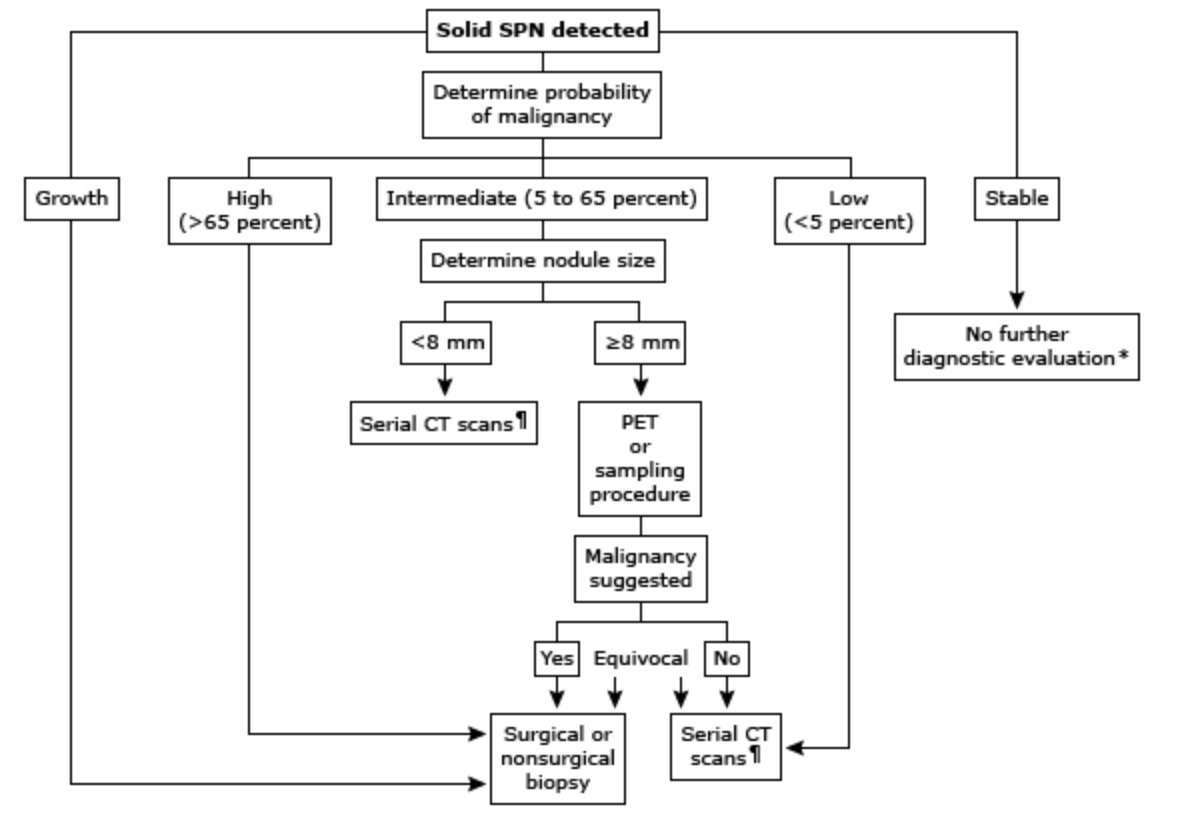
57yo man is referred to your practice due to an incidental 1.1cm single pulmonary nodule found on computed tomography. He is a never smoker and denies any known family history of lung cancer. He has no pulmonary medical history and reports no pulmonary symptoms.
What is the next step in the management of this patient?
Definition – wedge-shaped opacity in the periphery of the lung on chest radiography usually with its base along the pleural margins.
Clinical Significance – Occurs as a result of infarction and subsequent hemorrhage from the bronchial arteries classically due to a pulmonary embolism, but can also be from anything that causes infarction of lung parenchyma. The sensitivity and specificity of this finding is not robust and is, by definition, a late finding that is really no longer seen in modern medicine.
History –Named after Aubrey Otis Hampton (1900-1955), an American radiologist who received his medical degree from Baylor University in 1925. He rose through the ranks quickly in the field of radiology ultimately taking a position as chief of radiology at Massachussetts General in 1941. He first described his eponymous finding in 1940 in his manuscript entitled “Correlation of postmortem chest teleroentgenograms with autopsy findings”.
References
Firkin BG and Whitwirth JA. Dictionary of Medical Eponyms. 2nd ed. New York, NY; Parthenon Publishing Group. 1996.
Bartolucci S, Forbis P. Stedman’s Medical Eponyms. 2nd ed. Baltimore, MD; LWW. 2005.
Yee AJ, Pfiffner P. (2012). Medical Eponyms (Version 1.4.2) [Mobile Application Software]. Retrieved http://itunes.apple.com.
Schatzki R, Lingley JR. Aubrey O. Hampton, 1900-1955. The American journal of roentgenology, radium therapy, and nuclear medicine. 1956; 75(2):396-7. [pubmed]
Ladeiras-Lopes R, Neto A, Costa C, et al. Hampton’s hump and Palla’s sign in pulmonary embolism. Circulation. 2013; 127(18):1914-5. [pubmed]
Hampton AO, Castleman B. Correlation of postmortem chest teleroentogenograms with autopsy findings. Am J Roentgenol Radium Ther. 1940;34:305-326.
Wes Johnson, MSPAS, PA-C, (soon to be), DHSc was a former student of mine at UAB and was a respiratory therapist prior to PA school. He is the Regional Director of Clinical Education for Island Medical Management Emergency group in North Alabama. He won the Preceptor of The Year award from UAB in 2016 and currently finishing up his doctorate degree from A.T. Still University.
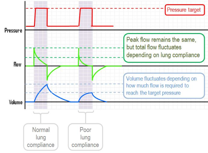
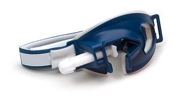
For the purposes of this podcast and post, we will be using the Puritan Bennett 840 ventilator (pictured below). All the term we use are synonymous with all vents, but the screens will be different.
Puritan Bennett 840
Big Concepts of The Ventilator
Mode
Assist Control (AC)
Every breath is either a machine driven (set by rate) or fully assisted (initiated by the patient)
Tobin MJ. Extubation and the myth of “minimal ventilator settings”. American journal of respiratory and critical care medicine. 2012; 185(4):349-50. [pubmed]
When following low-risk, small single pulmonary nodules,
What is the time sequence for the follow-up CT scans?
How long do you re-image them for?
Answers
It depends on the size of the nodule and consistency after the initial CT scan. The current guidelines state:
For solid nodules:
< 6mm recommend no further imaging
> 6mm but < 8mm recommend CT scan at 6-12 months
> 8mm recommend CT scan at 3-6 months, 9-12 months, and 18-24 months
For non-solid (ground glass) nodules:
< 5mm recommend no further imaging
> 5mm recommend annual CT for 3 years
For part-solid (>50% ground glass) nodules:
< 8mm recommend CT scan at 3 months, 12 months, 24 months, and annual for 1-3 years
> 8mm recommend CT scan at 3 months and PET or biopsy
Low-risk nodules can be serially scanned for 2 years and if no change, can stop repeat imaging as malignant nodules will show some form of change in 2 years.
References
Gould MK, Donington J, Lynch WR. Evaluation of individuals with pulmonary nodules: when is it lung cancer? Diagnosis and management of lung cancer, 3rd ed: American College of Chest Physicians evidence-based clinical practice guidelines. Chest. 2013; 143(5 Suppl):e93S-e120S. [pubmed]
300,000-600,000 cases per year in the United States
It is estimated that up to 50% will have post-thrombotic syndrome
Why Are We So Scared?
As many as 20% of patients with 1st onset PTE have no identifiable risk factors
10-30% 1-month mortality with up to 25% presenting as sudden death
Fear of litigation is #1 reason clinicians work-up low risk PTE
Why Can’t We Test Everyone?
Up to $16,000 per patient in total health care costs
6 times more deaths with testing and treatment
Signs and Symptoms
The majority of the classic signs and symptoms come from PIOPED II study and EMPEROR registry. These include:
Dyspnea (73%)
Chest Pain (64%)
Tachypnea (57%)
DVT findings or leg pain/swelling (47%)
Tachycardia (26%)
Dizziness (12%)
Hemoptysis (10%)
The EMPEROR registry took it a step further and determined mean vital sign measurements of:
Heart rate – 95 bpm
Respiratory rate – 20 bpm
Oxygen saturation – 95%
A recent trail in the NEJM called PESIT concluded as many as 1 in 6 patients with first time syncope has a PTE on inpatient work-up. This study has been largely panned by the EM community and you can read their take from the links below:
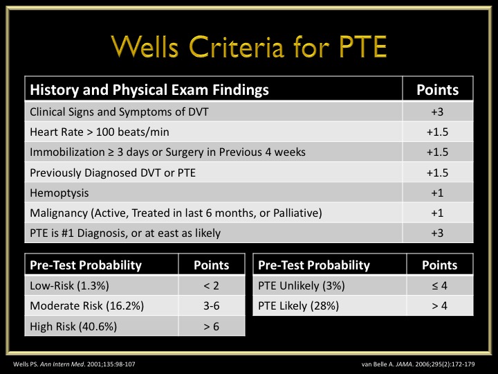
The most well know score is the Wells Criteria first published in 1998 and then revised and simplified in 2000 and 2001.
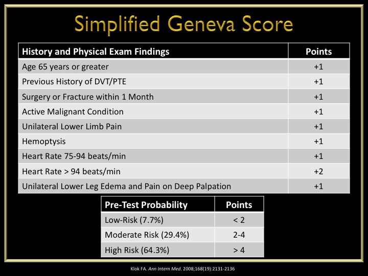
A second calculation is the Geneva Score first published in 2001 and revised and simplified in 2006 and 2008.
The Pulmonary Embolism Rule-Out Criteria was published in 2008 by Jeff Kline and is a second set of criteria to definitively rule-out PTE in patients ALREADY SCORE AS LOW RISK by Wells or Geneva.
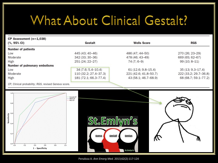
What about just good ol’ clinical gestalt? An interesting study was performed in 2013 looking at the accuracy of Wells vs Geneva vs Gestalt and found:
Clinical gestalt had a lower missed rate of PTE in low-risk patients
Clinical gestalt had a high accuracy of diagnosing PTE in high-risk patients
The Work-Up of Suspected PTE
Electrocardiogram is not senstitive nor specific for PTE but should be ordered on every patient with chest pain and/or shortness of breath to rule-out ACS
The EMCMD talks about the 10 ECG findings of PTE in the best video I have every scene
D-Dimer
High senstivity = good for rule-out
Should only be used after pre-test probability due to the false positives and unnecessary work-ups
ADJUST-PE Study
Found D-Dimer go up with age and created an age adjusted D-Dimer cutoff of:
Age (yr) x 10 as diagnostic threshold
Radiographic Imaging
Computed tomography is gold standard but has higher radiation exposure and contrast loads
Ventilation/Perfusion scan is safer in renal patients but up to 2/3rd are non-diagnostic
Risk Assessment
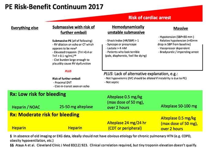
Once you diagnose a patient with a PTE, you have determine the patient’s risk and severity of disease.
Echocardiogram
Looking for RV strain
RV:LV ≥ 1
RV hypokinesis
Paradoxical septal movement
Tricuspid regurgitation
Biomarkers
Brain Natriuretic Peptide (BNP)
> 90 pg/mL has been associated with increased mortality
Troponin
> 0.01 ng/mL suggests evidence of RV dysfunction
Pulmonary Embolism Severity Index (PESI)
Published in 2005 and simplified 2010
Developed to help prognosticate 30d mortality and found low-risk patients (PESI – 0) can be safely treated as outpatient
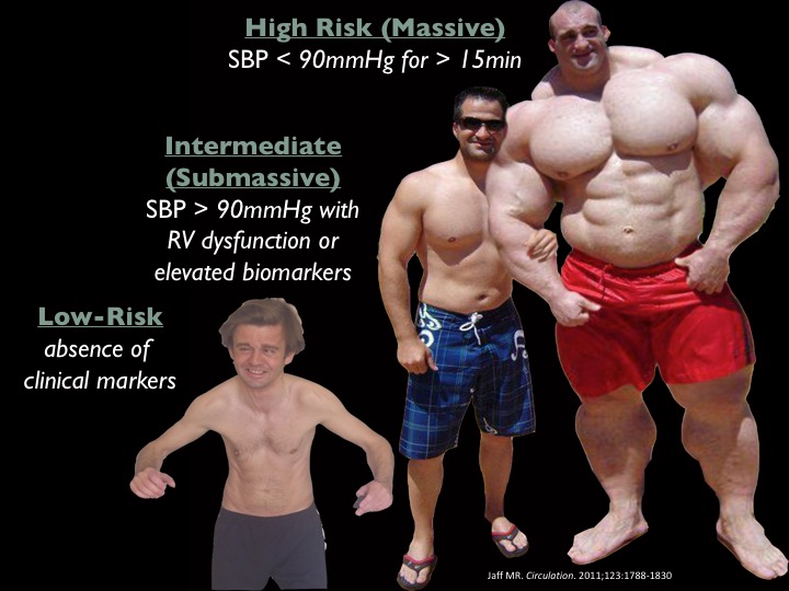
Definitions/Grades of PTE
Treatment Strategies for PTE
Anticoagulation
Started with confirmation of PTE or with high pre-test probability during workup
Lots of options (heparin, LMWH, direct thrombin inhibitors, Factor Xa inhibitors)
Fibrinolytics
Lots of recent research on who to lyse and who not to
Original research showed benefit if full dose lytics were given to massive PTE, but harm in submassive patients
This led to MOPPET in 2013 evaluating 1/2 dose lytics in submassive patients and found:
Reduction in overall mortality
No difference in bleeding complications
Reduction in hospital stay
PEITHO came next in 2014 and looked at full dose lytics vs anticoagulation only for submassive PTE and found:
No mortality benefit
Reduction in hemodynamic compromise
Increase in major bleeding and intracranial hemorrhage
Catheter Directed Therapy
Good options in patients with a high risk of bleeding with systemic fibrinolytic therapy
ULTIMA and SEATTLE-II studies found reduction in RV:LV ratio and decreased bleeding complications
Surgical Thrombectomy
Can be used as a last resort option and mortality from these procedures has dramatically improved from 57% in the 1960s to < 6% in 2005
Putting It All Together
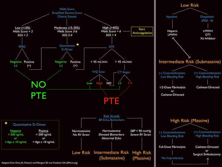
This is a graphic I modified from Jeff Kline and EmCrit that encompasses everything into a nice, neat package and as I have said before “algorithms will set you free”
References
Beckman MG. Venous Thromboembolism: A Public Health Concern. Am J Prev Med. 2010;38(4S):S495-S501
Spyropoulous AC. Direct medical costs of venous thromboembolism and subsequent hospital readmission rates: an administrative claims analysis from 30 managed care organizations. J Manag Care Pharm. 2007;13(6):475-486
Calder KK. The mortality of untreated pulmonary embolism in emergency department patients. Ann Emerg Med. 2005;45(3):302-310
Stein PD. Silent pulmonary embolism in patients with deep venous thrombosis: A systematic review. Am J Med. 2010;123:426-431
Stein PD. Clinical Characteristics of patients with acute pulmonary embolism. Am J Med. 2007;120:871-879
Pollack CV. Clinical characteristics, management, and outcoms of patients diagnosed with acute pulmonary embolism in the emergency department. JACC. 2011;57(6):700-706
Wells PS. Use of a clinical model for safe management of patients with suspected pulmonary embolism. Ann Intern Med. 1998;129:997-1005
Wells PS. Derivation of a simple clinical model to categorize patients probability of pulmonary embolism: increasing the models utility with the SimpliRED D-dimer. Thromb Haemost. 2000;83:416-420
Wells PS. Excluding pulmonary embolism at the bedside without diagbnostic imaging: management of patients with suspected pulmonary embolism presenting to the emergency department by using a simple clinical model and D-dimer. Ann Intern Med. 2001;135:98-107
van Belle A. Effectiveness of managing suspected pulmonary embolism using an algorithm combining clinical probability, D-dimer testing, and computed tomography. JAMA. 2006;295(2):172-179
Wicki J. Assessing clinical probability of pulmonary embolism in the emergency ward: a simple score. Arch Intern Med. 2001;161:997-92-97
Le Gal G. Prediction of pulmonary embolism in the emergency department: the revised Geneve score. Ann Intern Med. 2006;144:165-171
Klok FA. Simplication fo the revised Geneva score for assessing clinical probability of pulmonary embolism. Ann Intern Med. 2008;168(19):2131-2136
Kline JA. Prospective multicenter evaluation of the pulmonary embolism rule-out criteria. J Thromb Haemost. 2008;6:772-780
Penaloza A. Comparison of the unstructured clinical gestalt, the wells score, and the revised Geneva score to estimate pretest probability for suspected pulmonary embolism. Ann Emerg Med. 2013;62(2):117-124
Righini M. Age-adjusted D-dimer cutoff levels to rule-out pulmonary embolism: the ADJUST-PE study. 2014;311(11):1117-1124
Stein PD. Clinical characteristics of patients with acute pulmonary embolism: data from IOPED II. Am J Med. 2007;120:871-879
Anderson DR. Computerized tomographic pulmonary angiography versus ventilation perfusion lung scanning for the diagnosis of pulmonary embolism. Curr Opin Pulm Med. 2009;15:425–429
Rudoni RR. Use of two-dimensional echocardiography for the diagnosi of pulmonary embolus. J Emerg Med. 1998;16(1):5-8
Taylor RA. Point-of-care focused cardiac ultrasound for prediction of pulmonary embolism adverse outcomes. J Emerg Med. 2013;45(3):392-399
Kiely DG. Elevated levels of natriuretic peptides in patients with pulmonary thromboembolism. Resp Med. 2005;99:1286-1291
Jaff MR. Management of massive and submassive pulmonary embolism, iliofemoral deep vein thrombosis, chronic thromboembolic pulmonary hypertension: a scientific statement form the American Heart Association. Circulation. 2011;123:1788-1830
Keller K. Cardiac troponin I for predicting right ventricular dysfunction and intermediate risk in patients with normotensive pulmonary embolism. Neth Heart J. 2015;23:55-61
Aujesky D. Derivation and validation of a prognostic model for pulmonary embolism. Am J Respir Crit Care Med. 2005;172:1041-1046
Jimenez D. Simplification of the pulmonary embolism severity index for prognostication in patients with acute symptomatic pulmonary embolism. Arch Intern Med. 2010;170(15):1383-1389
Tapson VF. Treatment of pulmonary embolism: anticoagnulation, thrmbolytic therapy, and complications of therapy. Crit Care Clin. 2011;27:825-839
Sharifi M. Moderate pulmonary embolism treated with thrombolysis. Am J Cardiol. 2013;111:273-277
Zhang Z. Lower dosage of recombinant tissue-type plasminogen activator (rt-PA) in the treatment of acute pulmonary embolism: a systematic review and meta-analysis. Thrombosis Research. 2014;133:357-363
Meyer GM. Fibrinolysis for patients with intermediate-risk pulmonary embolism. NEJM. 2014;370(15):1402-1411
Chatterjee S. Thrombolysis for pulmonary embolism and risk of all-cause mortality, major bleeding, and intracranial hemorrhage: a meta-analysis. JAMA. 2014;311(23):2414-2421
Curtis GM. Risk factors associated with bleeding after alteplase administration for pulmonary embolism: a case control study. 2014;34(8):818–825
Kennedy RJ. Thrombus resolution and hemodynamic recovery using ultrasound-accelerated thrombolysis in acute pulmonary embolism. J Vasc Interv Radiol. 2013;24:841-848
Kucher N. Randomized, controlled trial of ultrasound-assisted catheter-directed thrombolysis for acute intermediate-risk pulmonary pulmonary embolism. Circulation. 2014;129:479-486
Cross FS. A survey of the current status of pulmonary embolectomy for massive pulmonary embolism. Circulation. 1967;35:186-191
Stulz P. Decision making in the surgical treatment of massive pulmonary embolism. Eur J Cardio-thorac Surg. 1994;8:188-193
Leacche M. Modern surgical treatment of massive pulmonary embolism: result in 47 sonsecutive patients after rapid diagnosis and aggressive surgical approach. J Thorac Cardiovasc Surg. 2005;129:1018-1023
Prandoni P, et al (PESIT Investigators). Prevalance of Pulmonary Embolism amount Patients Hospitalized for Syncope. NEJM. 2016;375:1524-1531
John MA, Klok FA, van Es N. D-dimer Interval Likelihood Ratios for Pulmonary Embolism. Acad Emerg Med. 2017;4;1-5.
Konstantinides SV, et al. Impact of Thrombolytic Therapy on the Long-Term Outcome of Intermediate-Risk Pulmonary Embolism. JACC. 2017;69(12):1536-1544
Sharifi M, et al (PEAPETT Investigators). Pulseless electrical activity in pulmonary embolism treated with thrombolysis. Am J Emerg Med. 2016;34(10):1963-1967
Piazza G, et al. (SEATTLE-II Investigators). A prospective, single arm, multicenter trial of ultrasound-facilitated , catheter-directed, low-dose fibrinolysis for acute massive and submassive pulmonary embolism. JACC. 2015;8(1):1382-1392
Definition – Form of hyperpnea (hyperventilation) characterized by a rhythmic, labored, and deep respiration pattern
Clinical Significance – Compensatory mechanism of profound metabolic acidosis, classically associated with diabetic ketoacidosis
History – Named after Adolph Kussmaul (1822-1902), a German physician, who noticed it in patients with severe diabetes mellitus and first published the finding in 1874. Dr. Kussmaul was a prolific physician in the late 1880’s and this is just one of many eponymous distinctions that bears his name. I am sure his name will come again in this series.
References
Firkin BG and Whitwirth JA. Dictionary of Medical Eponyms. 2nd ed. New York, NY; Parthenon Publishing Group. 1996.
Bartolucci S, Forbis P. Stedman’s Medical Eponyms. 2nd ed. Baltimore, MD; LWW. 2005.
Yee AJ, Pfiffner P. (2012). Medical Eponyms (Version 1.4.2) [Mobile Application Software]. Retrieved http://itunes.apple.com.
A. Kussmaul: Zur Lehre vom Diabetes mellitus. Über eine eigenthümliche Todesart bei Diabetischen, über Acetonämie, Glycerin-Behandlung des Diabetes und Einspritzungen von Diastase in’s Blut bei dieser Krankheit., Deutsches Archiv für klinische Medicin, Leipzig, 1874, 14: 1-46.
Young P, Finn BC, Bruetman JE, Buzzi A, Zylberman M. [The outstanding achievements of Adolf Kussmaul]. Revista medica de Chile. 2012; 140(4):538-44. [pubmed]
What are 3 pretest probability scoring systems used to evaluate patients with a suspected pulmonary thromboembolism?
Answer
There are 3 validated pretest probability scoring systems that can be used to help clinicians decide who can be sent home, who needs a D-dimer, and who goes straight to CT for suspected PTE.
Wells Criteria
Developed – 1998
Revised – 2000
Simplified – 2001
Geneva Score
Developed – 2001
Revised – 2006
Simplified – 2008
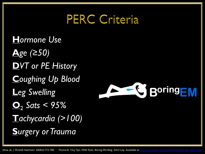
Pulmonary Embolism Rule-Out Criteria (PERC) Score
Developed – 2008
This score is used AFTER the patient is determined to be low-risk using the Well’s or Geneva score. In patients who are low-risk and PERC negative, there is only a 1.6% false-negative rate for missed PTE. Any one of these would deem the patient PERC positive.
Why is this so important?
Although it does help us in deciding who maybe at higher risk of PTE, I personally feel these scoring systems help us document who DOES NOT need work-up. There are quite a few patients who come in with non-specific chest pain or shortness of breath, and you should ALWAYS entertain the idea of PTE in these patients. But, not every single one of these patients need a d-dimer or CTA. Better yet, some of these patients can be discharged home without any investigation if they are low-risk and PERC negative.
Below is an algorithm I modified from Jeff Kline using these clinical decision instruments.
All these images are slides from my talk at the 2015 AAPA Conference
References
Wells PS, Ginsberg JS, Anderson DR. Use of a clinical model for safe management of patients with suspected pulmonary embolism. Annals of Internal Medicine. 1998;129(12):997-1005. [pubmed]
Wells PS, Anderson DR, Rodger M. Derivation of a simple clinical model to categorize patients probability of pulmonary embolism: increasing the models utility with the SimpliRED D-dimer. Thrombosis and Haemostasis. 2000;83(3):416-20. [pubmed]
Wells PS, Anderson DR, Rodger M. Excluding pulmonary embolism at the bedside without diagnostic imaging: management of patients with suspected pulmonary embolism presenting to the emergency department by using a simple clinical model and d-dimer. Annals of Internal Medicine. 2001;135(2):98-107. [pubmed]
Wicki J, Perneger TV, Junod AF, Bounameaux H, Perrier A. Assessing clinical probability of pulmonary embolism in the emergency ward: a simple score. Archives of Internal Medicine. 2001;161(1):92-7. [pubmed]
Le Gal G, Righini M, Roy PM. Prediction of pulmonary embolism in the emergency department: the revised Geneva score. Annals of Internal Medicine. 2006;144(3):165-71. [pubmed]
Klok FA, Mos IC, Nijkeuter M. Simplification of the revised Geneva score for assessing clinical probability of pulmonary embolism. Archives of Internal Medicine. 2008;168(19):2131-6. [pubmed]
Kline JA, Courtney DM, Kabrhel C. Prospective multicenter evaluation of the pulmonary embolism rule-out criteria. Journal of Thrombosis and Haemostasis. 2008; 6(5):772-80. [pubmed]
This is actually a special episode for the PAINE Podcast as I have the opportunity to do a joint-interview podcast with Chip Lange from TOTAL EM. This was the first time I got to dabble with a conversational-style podcast and I think it went pretty good. Chip and I had a great time doing it and will most definitely be doing more of these in the future.
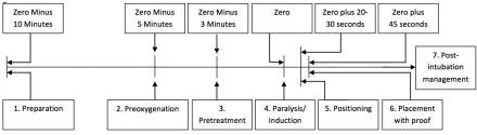
One of the many saying my Army Airborne Ranger dad has instilled in me growing (and one that I still use today) is the seven “P” approach to accomplishing tasks:
Proper
Planning
and
Preparation
Prevents
Piss
Poor
Performance
What is nice about this saying is that it applies very nicely to the steps of intubation as well.
Prepare
You need to to have everything at the bedside you MIGHT need prior to any intubation attempt. This includes equipment, medications, and any personnel or team members who will assist. If you even suspect this could be a difficult airway, you should have your plan B and plan C options in the room to ward off the evil spirits.
If using video, plug it in and make sure it turns on
Patent IV lines x 2
Suction
Cardiac and pulse oximetry monitor
Bag-valve mask
End-tidal CO2 monitor
Medications
Drawn up and labeled
Concentration read aloud
This also gives you the opportunity to talk with you team about the plan for intubation (how many attempts, progression should plan A, steps of what will happen during the intubation and everyone’s roles during the procedure, etc..), as well as reviewing assisting maneuvers (external laryngeal manipulation, etc.).
Preoxygenate
In order to decrease any deoxygenation-related issues during the intubation attempt, your patient should recieve 100% oxygen at 15 liters per minute through a non-rebreather mask for 3-5 minutes. This will properly de-nitrogenate and super-saturate all the hemoglobin and give you the time you need to visualize and intubate.
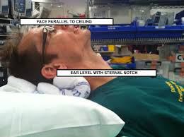
Position
“EAR HOLE TO CHEST HOLE”
For ideal visualization, you want to position your patient so that their external auditory meatus lined up to the sternal notch
Premedicate
There are several different medications you can give for premedication purposes to modify the physiologic response during intubation (lidocaine, opiates, atropine, defasculating agents, etc..), but the main one is the sedative. It is generally poor form to paralyze someone before you sedate them. There are several medications you can choose from for sedation in intubation:
Ketamine – 1-2mg/kg IV
My ideal sedative
Etomidate 0.3mg/kg IV
Less hemodynamic compromise
Can cause adrenal suppression
Propofol – 1.5-3mg/kg IV
Can cause hemodynamic instability
Paralyze
There are 2 choices for classes of paralytics:
Depolarizing
Succinylcholine – 1.5-2mg/kg IV
Rapid onset, shortest duration of action
Caution in burn/crush injuries, hyperkalemic patients
Non-Depolarizing
Rocuronium – 1.2mg/kg IV
Vecuronium – 0.3mg/kg IV
Pass The Tube
Once you patient is properly sedative and paralyze, you can proceed to laryngoscopy.
Post-intubation Assessment
Capnography
This is used for confirmation of correct placement of the endotracheal in the trachea and tests for end-tidal CO2. There are 2 main types:
Qualitative
Color change calorimeter
Attaches to end of endotracheal tube and detects CO2 by changes in exhaled pH
GOLD IS GOOD
Quantitative
Continuous Waveform Capnography
Gold standard
Gives you a visual waveform to see if the ventilations are adequate
Securing the Tube
Once you know you are in the right spot and have been confirmed by capnography, you need to secure the tube. There are different ways to achieve and I often defer to the respiratory therapist or nurse on how they want it secured. There are commercial devices that lock the tube in place and secure using velcro straps, all the way to the old standby of adhesive tape. This is a great site that shows several different ways you can secure the endotracheal tube (http://aam.ucsf.edu/article/securing-endotracheal-tube).
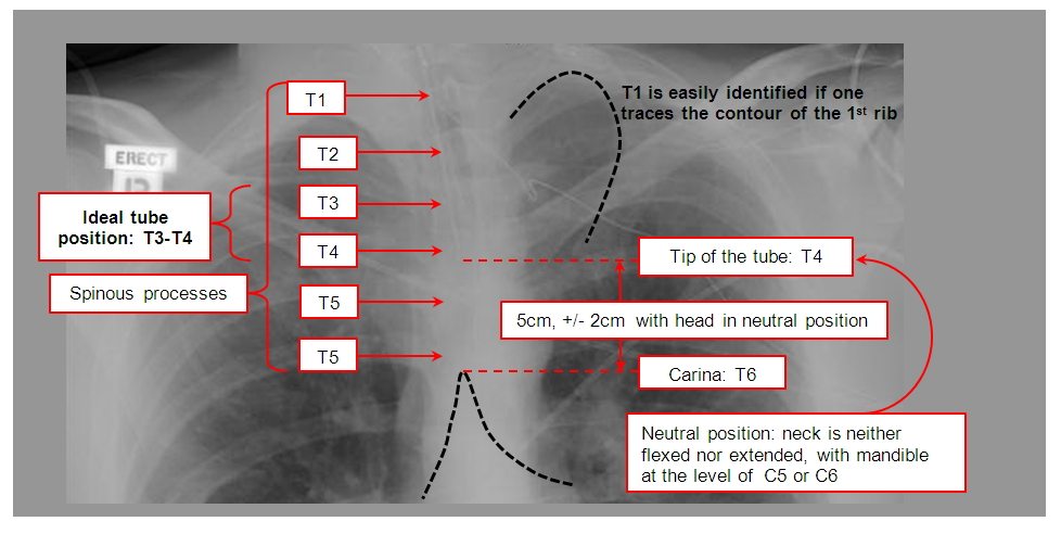
Radiography
Chest xray is the gold standard for the radiographical confirmation of endotracheal placement, as well as ensuring the proper depth. The ideal position for the tube depth should be 3-5cm from the carina or at T3-4 position.
Now that the tube is in place, secured, and confirmed, you are done right? WRONG!!! Your patient now has a tube shoved into the tracheal and it is a tad uncomfortable. Postintubation sedation/analgesia is PARAMOUNT for good patient care.
Sedation
Ketamine – 0.1-0.5mg/kg bolus and 0.1-0.5mg/kg/hr infusion
Propofol – 5mcg/kg bolus and 5-50mcg/kg/hr infusion
Midazolam – 0.05mg/kg bolus and 0.025mg/kg/hr infusion
Analgesia
Fentanyl – 2mcg/kg bolus and 1mcg/kg/hr infusion
Hydromorphone – 0.5-1mg/kg bolus and 0.5-3mg/kg/hr infusion
Morphine – 5-10mg/kg bolus and 2-30mg/hr infusion
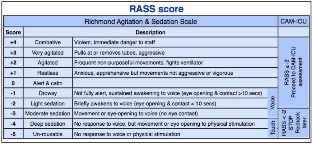
You should be shooting for a Richmond Agitation Sedation Scale (RASS) of -1 to -3 for adequate sedation following intubation.