Ep-PAINE-nym


Jones Fracture


Other Known Aliasesnone


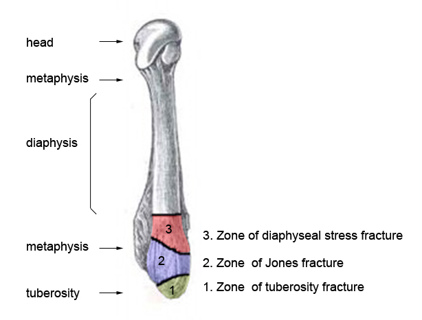
Definition – fracture of the base of the 5th metatarsal at the metaphyseal-diaphyseal junction
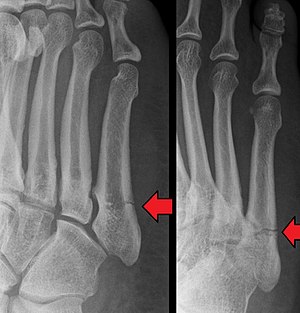
Image result for jones fracture

Clinical SignificanceFairly easy radiographical diagnosis to make, but careful examination must distinguish between acute injuries in Zone 1 and 2 and chronic injuries in Zone 3.

Image result for jones fractureImage result for jones fracture zones

History – Named after Sir Robert Jones (1857-1933), a Welsh orthopaedic surgeon and received his medical doctorate from the Liverpool School of Medicine in 1887 and achieved fellowship in 1889.  Along with his uncle, he was a pioneer in the diagnosis and management of fractures.  He first described the injury that bears his name in 1902 in the Annals of Surgery entitled “Fracture of the Base of the Fifth Metatarsal by Indirect Violence”.  This paper was a six patient case report on the injury pattern and Dr. Jones was patient number one having injured his foot several months prior dancing.  After Wilhem Rontgen published his discovery of x-rays in 1895, Dr. Jones adopted this new modality fully in the practice of orthopaedics and published the first clinical radiograph in 1896 about a 12yo with a bullet lodged in his wrist that could not be found clinically and required a 2hr long exposure.

Robert Jones (surgeon).jpg

scanned image of page 697


References

  1. Firkin BG and Whitwirth JA.  Dictionary of Medical Eponyms. 2nd ed.  New York, NY; Parthenon Publishing Group. 1996.
  2. Bartolucci S, Forbis P.  Stedman’s Medical Eponyms.  2nd ed.  Baltimore, MD; LWW.  2005.
  3. Yee AJ, Pfiffner P. (2012).  Medical Eponyms (Version 1.4.2) [Mobile Application Software].  Retrieved http://itunes.apple.com.
  4. Whonamedit – dictionary of medical eponyms. http://www.whonamedit.com
  5. Up To Date. www.uptodate.com
  6. Jones Fracture.  Wheeless’ Textbook of Orthopaedics. http://www.wheelessonline.com/ortho/jones_fracture
  7. OrthoBullets. https://www.orthobullets.com/foot-and-ankle/7031/5th-metatarsal-base-fracture
  8. Jones R. I. Fracture of the Base of the Fifth Metatarsal Bone by Indirect Violence. Annals of surgery. 1902; 35(6):697-700.2. [pubmed]
  9. Jones R, Lodge O.  The Discovery of a Bullet Lost in the Wrist by Means of the Roentgen Rays.  Lancer. 1896;147(3782):476-477 [article]

PAINE #PANCE Pearl – Infectious Disease


Question


To weave a common theme this week between this post and Wednesday’s eponym:

  1. What are the HACEK bugs?
  2. What disease are they associate with?
  3. Why are they important?


Answer


The HACEK pathogens is an acronym for the fastidious, gram-negative bacteria that are implicated in 5-10% of infective endocarditis cases. The bugs are:

  • Haemophilus species
  • Aggregatibacter species
  • Cardiobacterium species
  • Eikenella species
  • Kingella species


These pathogens are normal oropharyngeal flora, but can take up to 14 days to grow in the laboratory and are often referred to as
culture-negative” endocarditis cases.  It is important to discuss with your lab if you are worried about HACEK pathogens so they can plate the blood cultures on the appropriate agar plates and keep past the typical 3-5 days if there is no growth.


References

  1. Sharara SL, Tayyar R, Kanafani ZA, Kanj SS. HACEK endocarditis: a review. Expert review of anti-infective therapy. 2016; 14(6):539-45. [pubmed]
  2. Chambers ST, Murdoch D, Morris A, et al. HACEK infective endocarditis: characteristics and outcomes from a large, multi-national cohort. PloS one. 2013; 8(5):e63181. [pubmed]
  3. Yew HS, Chambers ST, Roberts SA, et al. Association between HACEK bacteraemia and endocarditis. Journal of medical microbiology. 2014; 63(Pt 6):892-5. [pubmed]
  4. Wassef N, Rizkalla E, Shaukat N, Sluka M. HACEK-induced endocarditis. BMJ case reports. 2013; 2013:. [pubmed]

Ep-PAINE-nym


Duke Criteria


Other Known Aliasesnone


Definitionradiographic, laboratory, pathologic, and clinical criteria to help establish the diagnosis of infective endocarditis and (similar to the Jones Criteria) there are major and minor subtypes:

  • Major
    • Two positive blood cultures (drawn 12 hours apart) with typical endocarditis pathogen
      • Viridans-group streptococci
      • Streptococcus bovis
      • HACEK group
      • Staphylococcus aureus
      • Community-acquired enterococci
      • Coxiella burnetii
    • Evidence of endocardial involvement with positive echocardiogram showing:
      • oscillating intracardiac mass
      • Abscess
      • partial dehiscence of prosthetic valve or new valvular regurgitation
  • Minor
    • known cardiac lesion or IVDU
    • Fever > 38°C
    • Evidence of septic emboli
    • Immunologic phenomenon: glomerulonephritis, Osler’s nodes, Janeway lesions, conjunctival hemorrhage
    • Positive blood culture  or laboratory evidence of organisms not described above


Clinical Significancea diagnosis of infective endocarditis is made with 1 major and 1 minor, or 3 minor criteria


History – Named after the Duke Endocarditis Service, which established these criteria with their seminal paper entitled “New Criteria for the diagnosis of infective endocarditis” in 1994.  The group sought to improve upon the older von Reyn criteria and increase the specificity, so as to not miss cases and have potentially catastrophic cardiac complications later.  These criteria were further validated several times throughout the 1990s and modified in 2000, which is the current set we use today.


References

  1. Firkin BG and Whitwirth JA.  Dictionary of Medical Eponyms. 2nd ed.  New York, NY; Parthenon Publishing Group. 1996.
  2. Bartolucci S, Forbis P.  Stedman’s Medical Eponyms.  2nd ed.  Baltimore, MD; LWW.  2005.
  3. Yee AJ, Pfiffner P. (2012).  Medical Eponyms (Version 1.4.2) [Mobile Application Software].  Retrieved http://itunes.apple.com.
  4. Whonamedit – dictionary of medical eponyms. http://www.whonamedit.com
  5. Up To Date. www.uptodate.com
  6. Durack DT, Lukes AS, Bright DK. New criteria for diagnosis of infective endocarditis: utilization of specific echocardiographic findings. Duke Endocarditis Service. Am J Med. 1994; 96(3):200-9. [pubmed]
  7. Li JS, Sexton DJ, Mick N, et al. Proposed modifications to the Duke criteria for the diagnosis of infective endocarditis. Clinical infectious diseases : an official publication of the Infectious Diseases Society of America. 2000; 30(4):633-8. [pubmed]

Ep-PAINE-nym


Jones Criteria


Other Known Aliasesdiagnostic criteria for acute rheumatic fever


Definitionclinical criteria to help diagnose acute rheumatic fever.  There are divided into major and minor criteria as follows:

  • Major
    • Polyarthritis
    • Carditis
    • Subcutaneous nodules
    • Erythema marginatum
    • Sydenham’s chorea
  • Minor
    • Fever
    • Arthralgia
    • Elevated ESR or CRP
    • Leukocytosis
    • 1st degree heart block

Clinical Significancea diagnosis of acute rheumatic fever is either two major or one major and two minor criteria


History – Named after T. Duckett Jones (1899-1954), an American cardiologist who received his medical doctorate from the University of Virginia in 1923.  With a keen interest in rheumatic fever and heart disease, he practiced at Massachusetts General Hospital and House of Good Samaritan in Boston for over 20 years.  He became the medical director of the Helen Way Whitney Foundation to pursue his passion for public health, which led to one of the first tweleve appointments to the National Advisory Heart Council in 1948.  He published his seminal paper entitled “The Diagnosis of Rheumatic Fever” in JAMA in 1944 which described these findings.  Dr. Jones unfortunately died as a result of malignant hypertension in 1954 at the age of 55.

First page PDF preview


References

  1. Firkin BG and Whitwirth JA.  Dictionary of Medical Eponyms. 2nd ed.  New York, NY; Parthenon Publishing Group. 1996.
  2. Bartolucci S, Forbis P.  Stedman’s Medical Eponyms.  2nd ed.  Baltimore, MD; LWW.  2005.
  3. Yee AJ, Pfiffner P. (2012).  Medical Eponyms (Version 1.4.2) [Mobile Application Software].  Retrieved http://itunes.apple.com.
  4. Whonamedit – dictionary of medical eponyms. http://www.whonamedit.com
  5. Up To Date. www.uptodate.com
  6. White PD.  T. Duckett Jones, 1899-1954.  Circulation.  1955.
  7. Shulman ST. T. Duckett Jones and his criteria for the diagnosis of acute rheumatic fever. Pediatric annals. 1999; 28(1):9-12. [pubmed]
  8. Jones TD.  The Diagnosis of Rheumatic Fever.  JAMA. 1944;126(8):481-484 [article]

PAINE #PANCE Pearl – Pulmonary


Question


57yo man is referred to your practice due to an incidental 1.1cm single pulmonary nodule found on computed tomography.  He is a never smoker and denies any known family history of lung cancer.  He has no pulmonary medical history and reports no pulmonary symptoms.


Image result for single pulmonary nodule ct


What is the next step in the management of this patient?


Answer


  1. The first step in the management of this patient should be investigate if there are any previous studies to compare.  This will allow us to be able to assess if any growth has taken place.  For this patient, there are no previous studies to compare.
  2. The next step would be assess malignancy risk since it is larger than 8mm.  Up to Date uses the Brock University Cancer Equation, which I happen to like as well, though there are several out there and all use clinical, historical, and radiographical criteria.
  3. Our patient has a predicted malignancy risk of 3.54% and using the below algorithm, this patient needs a follow-up CT scan in 3 months to evaluate growth.


References

  1. Gould MK, Donington J, Lynch WR, et al. Evaluation of individuals with pulmonary nodules: when is it lung cancer? Diagnosis and management of lung cancer, 3rd ed: American College of Chest Physicians evidence-based clinical practice guidelines. Chest. 2013; 143(5 Suppl):e93S-e120S. [pubmed]
  2. Callister ME, Baldwin DR, Akram AR, et al. British Thoracic Society guidelines for the investigation and management of pulmonary nodules. Thorax. 2015; 70 Suppl 2:ii1-ii54. [pubmed]
  3. McWilliams A, Tammemagi MC, Mayo JR, et al. Probability of cancer in pulmonary nodules detected on first screening CT. The New England journal of medicine. 2013; 369(10):910-9. [pubmed]
  4. Up-To-Date.  Diagnostic Evaluation of the Incidental Pulmonary Nodule.  2018.

Ep-PAINE-nym


Westermark’s Sign


Other Known Aliasesnone


Definitionfocal peripheral hyperlucency resulting from collapsed vessels distal to a pulmonary thromboembolism.


Clinical SignificanceOccurs as a result of oligemia of perfusion to the lung parenchyma and can be seen in up to 10% of patients with acute PTE.  Similar to Hampton’s Hump, it has a low sensitivity, but a high specificity


History – Named after Nils Johan Hugo Westermark (1892-1980), a Swedish radiologist who first described this finding in his 1938 paper entitled ” On the roentgen diagnosis of lung embolism”.  He was also an accomplished sailor and won a silver medal in the 1912 Olympics.


References

  1. Firkin BG and Whitwirth JA.  Dictionary of Medical Eponyms. 2nd ed.  New York, NY; Parthenon Publishing Group. 1996.
  2. Bartolucci S, Forbis P.  Stedman’s Medical Eponyms.  2nd ed.  Baltimore, MD; LWW.  2005.
  3. Yee AJ, Pfiffner P. (2012).  Medical Eponyms (Version 1.4.2) [Mobile Application Software].  Retrieved http://itunes.apple.com.
  4. Whonamedit – dictionary of medical eponyms. http://www.whonamedit.com
  5. Up To Date. www.uptodate.com
  6. Krishnan AS, Barrett T. Images in clinical medicine. Westermark sign in pulmonary embolism. NEJM. 2012; 366(11):e16. [pubmed]
  7. Radiopaedia.  Westermark Sign. https://radiopaedia.org/articles/westermark-sign-1
  8. Westermark N. On the roentgen diagnosis of lung embolism. Acta Radiol 1938;19:357‑72.

PAINE #PANCE Pearl – Pulmonary


57yo man is referred to your practice due to an incidental 1.1cm single pulmonary nodule found on computed tomography.  He is a never smoker and denies any known family history of lung cancer.  He has no pulmonary medical history and reports no pulmonary symptoms.


Image result for single pulmonary nodule ct


What is the next step in the management of this patient?

Ep-PAINE-nym


Hampton’s Hump


Other Known Aliasesnone


Definitionwedge-shaped opacity in the periphery of the lung on chest radiography usually with its base along the pleural margins.


Clinical SignificanceOccurs as a result of infarction and subsequent hemorrhage from the bronchial arteries classically due to a pulmonary embolism, but can also be from anything that causes infarction of lung parenchyma.  The sensitivity and specificity of this finding is not robust and is, by definition, a late finding that is really no longer seen in modern medicine.


History – Named after Aubrey Otis Hampton (1900-1955), an American radiologist who received his medical degree from Baylor University in 1925.  He rose through the ranks quickly in the field of radiology ultimately taking a position as chief of radiology at Massachussetts General in 1941.  He first described his eponymous finding in 1940 in his manuscript entitled “Correlation of postmortem chest teleroentgenograms with autopsy findings”.

Image result for Aubrey Otis Hampton


References

  1. Firkin BG and Whitwirth JA.  Dictionary of Medical Eponyms. 2nd ed.  New York, NY; Parthenon Publishing Group. 1996.
  2. Bartolucci S, Forbis P.  Stedman’s Medical Eponyms.  2nd ed.  Baltimore, MD; LWW.  2005.
  3. Yee AJ, Pfiffner P. (2012).  Medical Eponyms (Version 1.4.2) [Mobile Application Software].  Retrieved http://itunes.apple.com.
  4. Whonamedit – dictionary of medical eponyms. http://www.whonamedit.com
  5. Up To Date. www.uptodate.com
  6. Radiopaedia. Hamptons’ Hump. https://radiopaedia.org/articles/hampton-hump-2
  7. Schatzki R, Lingley JR. Aubrey O. Hampton, 1900-1955. The American journal of roentgenology, radium therapy, and nuclear medicine. 1956; 75(2):396-7. [pubmed]
  8. Ladeiras-Lopes R, Neto A, Costa C, et al. Hampton’s hump and Palla’s sign in pulmonary embolism. Circulation. 2013; 127(18):1914-5. [pubmed]
  9. Hampton AO, Castleman B.  Correlation of postmortem chest teleroentogenograms with autopsy findings.  Am J Roentgenol Radium Ther. 1940;34:305-326.

PAINE #PANCE Pearl – Surgery


Question


What is the main difference between these two instruments?


Image result for allis clampImage result for allis clamp


vs


Image result for babcock clampImage result for babcock clamp


Answer


The first instrument is an Allis clamp, which has sharp teeth and can crush tissue.  It is used for grasping fascia or tissue that needs to be removed or manipulated during procedures.


The second instrument is a Babcock forceps,  which is non-toothed and has a wider grasping surface.  These forceps do not damage tissue and are considered non-crushing and can be used to grasp delicate tissue.  The head of the forceps is open and helps for rapid identification.