Ep-PAINE-nym


Naegele’s rule

Other Known Aliasesestimated date of delivery

Definitionestimation of delivery assuming a 280 day gestation period and is calculated from the FIRST day of the last menstrual cycle by adding 1 year, subtracting 3 months, and adding 7 days.

Clinical Significance this is a quick and easy estimation of the delivery date for planning purposes and is used in most apps and delivery wheels. In the age of ease of ultrasound, direct measurement is becoming the standard, but this is still a very important calculation to remember.
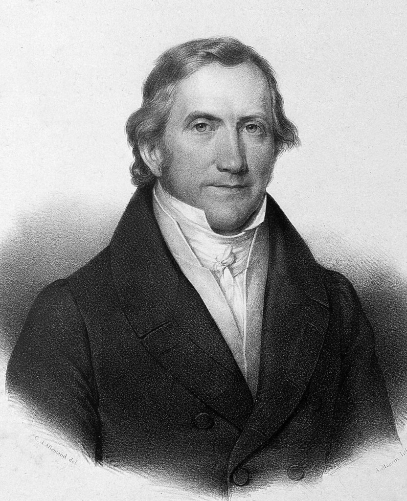
HistoryNamed after Franz Karl Naegele (1778-1851), who was a German obstetrician and received his medical doctorate from the the University of Bamberg. He had a very successful practice in Barmen, Germany, before he went on to become full professor of obstetrics in 1810 at the University of Heidelberg. He first mentioned his rule, and credited Hermann Boerhaave who first mentioned it in 1744, in a manuscript in 1812, but was given the eponym by Gunning Bedford, professor of obstetrics and diseases of Women and Children at the University of New York, in 1872.


References

  1. Firkin BG and Whitwirth JA.  Dictionary of Medical Eponyms. 2nd ed.  New York, NY; Parthenon Publishing Group. 1996.
  2. Bartolucci S, Forbis P.  Stedman’s Medical Eponyms.  2nd ed.  Baltimore, MD; LWW.  2005.
  3. Yee AJ, Pfiffner P. (2012).  Medical Eponyms (Version 1.4.2) [Mobile Application Software].  Retrieved http://itunes.apple.com.
  4. Whonamedit – dictionary of medical eponyms. http://www.whonamedit.com
  5. Up To Date. www.uptodate.com
  6. Baskett TF. Eponym and Names in Obstetrics and Gynaecology. 3rd Ed. Cambridge, UK. Cambridge University Press. 2019.
  7. Baskett TF, Nagele F. Naegele’s Rule: a reappraisal. BJOG. 200;107(1):1433-1435.
  8. Naegele FC. Erfahrungen und Abhandlungen aus dem Gebiethe der Krankheiten des Weiblichen Geschlechtes. Nebst Grundziigen einer Methodenlehre der Geburtshiilfe. Mannheim: Loeffler, 1812: 280-281
  9. Bedford GS. The Principles and Practice of Obstetrics. 5th Edition. New York William Wood and Co, 1872:306.

#56 – Polycystic Ovarian Syndrome


***LISTEN TO THE PODCAST HERE***


Background

  • First described by Stein and Leventhal in 1935
  • The most common cause of infertility in women
    • Up to 30% of women seeking infertility treatment
  • Affects 6-12% of US women ( or 1 in 10)  of reproductive age
  • Increases life-time risk of developing:
    • Obesity
    • DMII
    • Cardiovascular disease
    • Breast and endometrial cancers

Pathophysiology

  • Two-Hit Hypothesis
    • First – genetic predisposition
      • Heritable traits and gene variations affecting ovarian function, insulin resistance, obesity, and DMII
        • 25% of patients with PCOS have a mother with PCOS
      • Congenital virilization
        • Congenital adrenal hyperplasia
      • Disturbed fetal nutrition
    • Second – provocative trigger
      • Insulin-resistant hyperinsulinemia
      • Puberty
  • This then leads to the classic pathology of:
    • Functional ovarian hyperandrogenism
    • Hyperinsulinism and obesity
    • Luteinizing hormone (LH) excess
Up-to-Date

Definition and Diagnostic Criteria

  • Adults
    • Rotterdam Criteria
      • 2 of 3 following criteria:
        • Anovulation
        • Hyperandrogenism
        • Polycystic ovaries
Up-to-Date
  • Adolescents
    • Developed in 2015 and consist of otherwise unexplained persistent hyperandrogenic oligo-anovulatory menstrual abnormality based on age and stage appropriate standards
Up-to-Date

Clinical Features

  • Cutaneous Hyperandrogenism
    • Hirsutism
      • Graded by Ferriman-Gallwey scoring system, which quantitates the extent of hair growth in androgen sensitive areas
        • Hirsutism is defined as a score ≥ 8
    • Acne
      • Moderate comedonal acne or severe inflammatory acne suggests hyperandrogenemia
  • Ovarian Findings
    • Menstrual
      • Primary Amenorrhea
        • Lack of menarch by 15 years of age or > 3 years after onset of breast development
      • Secondary Amenorrhea
        • > 90 days without a menstrual cycle after previously menstruating
      • Oligomenorrhea
        • During the first five years after menarache:
          • Year 1 – < 4 cycles in the year
          • Year 2 – < 6 cycles in the year
          • Year 3-5 – < 8 cycles in the year
            • Missing ≥ 4 cycles in the year
          • Year 6+ – < 9 cycles in the year
            • Missing ≥ 3 ycles in the year
      • Excessive uterine bleeding
        • More frequently than every 21 days or excessive bleeding
          • PCOS is the most common cause of excessive uterine bleeding in adolescents
    • Polycystic ovaries
  • Obesity
    • Chief complaint in up to 20% of PCOS patients
  • Sleep apnea or
  • Nonalcoholic fatty liver
  • Manifestations of insulin resistance
    • Acanthosis nigricans
    • Metabolic syndrome
      • Up to 25% of PCOS patient

Diagnostic Work-Up

  • Need to be performed at a lab with highly sensitive assay capability
  • If using hormonal OCP, need to be stopped 2-3 months before testing
    • Due to suppression of testosterone
  • Testosterone (1st step)
    • Should be early morning as testosterone levels fall by the afternoon
    • Serum total testosterone
      • Normal – 40-60 ng/dL
      • > 150 ng/dL is diagnostic
    • Serum free testosterone
      • More sensitive than total, but are less standardized
      • Only reliable if calculated from the total testosterone
  • Endocrine Screening Panel (2nd step if elevated testosterone)
    • Beta-hCG
    • FSH/LH
      • Slightly elevated LH with a slightly decreased FSH is characteristic of PCOS
      • Markedly elevated FSH = primary hypogonadism
      • Markedly decreased LH = secondary hypogonadism
    • TSH
  • Screening for Common non-PCOS causes of hyperandrogenism (3rd step if endocrine screening is normal)
    • 17-hydroxyprogesterone (17OHP)
      • Drawn at 0800 and with the patient either amenorrheic or within the fist 10 days after the start of her menstrual cycle
      • > 170 ng/dL suggests CAH
    • DHEAS
      • > 700 mcg/dL suggests adrenal tumor
    • Prolactin
      • Hyperprolactinemia can causes gonadotropin deficiency
      • > 25 ng/m: suggests prolactinoma
    • Serum cortisol
      • < 10 mcg/dL rules out Cushing syndrome
    • Insulin-like grown factor (IGF-1)
      • Rule out acromegaly
  • Other tests
    • Chronic disease panel
      • CBC, ESR/CRP, CMP
    • Lipid Panel (for adults)
      • LDL, HDL, triglycerides
  • Transvaginal ultrasound of ovaries
    • Increased overall size
    • Increased number of distinct follicles
      • ≥ 6 is diagnostic

Treatment

  • Adolescents
    • Antiandrogen
      • Estrogen-progestin combination OCPs
        • Can also use GnRH agonist (leuprolide)
      • Targeted antiandrogen therapy (if no improvement after 6 months)
        • Spironolactone
        • Finasteride
    • Insulin resistance
      • Biguanide (metformin)
      • Thiazolidinediones (pioglitazone, rosiglitazone)
  • Adults
    • Same as above, but add:
      • Dyslipidemia therapy

The Cottage Physician (1893)


References

  1. Stein IF, Leventhal ML.  Amenorrhea associated with bilateral polycystic ovaries.  AJOG. 1935;29(2):181-191 [article]
  2. Azziz R, Woods KS, Reyna R, Key TJ, Knochenhauer ES, Yildiz BO. The prevalence and features of the polycystic ovary syndrome in an unselected population. The Journal of clinical endocrinology and metabolism. 2004; 89(6):2745-9. [pubmed]
  3. Franks S, Stark J, Hardy K. Follicle dynamics and anovulation in polycystic ovary syndrome. Human reproduction update. ; 14(4):367-78. [pubmed]
  4. Barthelmess EK, Naz RK. Polycystic ovary syndrome: current status and future perspective. Frontiers in bioscience (Elite edition). 2014; 6:104-19. [pubmed]
  5. Rotterdam ESHRE/ASRM-Sponsored PCOS Consensus Workshop Group. Revised 2003 consensus on diagnostic criteria and long-term health risks related to polycystic ovary syndrome. Fertility and sterility. 2004; 81(1):19-25. [pubmed]
  6. Azziz R, Carmina E, Dewailly D, et al. The Androgen Excess and PCOS Society criteria for the polycystic ovary syndrome: the complete task force report. Fertility and sterility. 2009; 91(2):456-88. [pubmed]
  7. Rosenfield RL. The Diagnosis of Polycystic Ovary Syndrome in Adolescents. Pediatrics. 2015; 136(6):1154-65. [pubmed]
  8. Witchel SF, Oberfield S, Rosenfield RL, et al. The Diagnosis of Polycystic Ovary Syndrome during Adolescence. Hormone research in paediatrics. 2015; [pubmed]
  9. Martin KA, Anderson RR, Chang RJ, et al. Evaluation and Treatment of Hirsutism in Premenopausal Women: An Endocrine Society Clinical Practice Guideline. The Journal of clinical endocrinology and metabolism. 2018; 103(4):1233-1257. [pubmed]
  10. Maslyanskaya S, Talib HJ, Northridge JL, Jacobs AM, Coble C, Coupey SM. Polycystic Ovary Syndrome: An Under-recognized Cause of Abnormal Uterine Bleeding in Adolescents Admitted to a Children’s Hospital. Journal of pediatric and adolescent gynecology. 2017; 30(3):349-355. [pubmed]
  11. Helvaci N, Karabulut E, Demir AU, Yildiz BO. Polycystic ovary syndrome and the risk of obstructive sleep apnea: a meta-analysis and review of the literature. Endocrine connections. 2017; 6(7):437-445. [pubmed]
  12. Elhassan YS, Idkowiak J, Smith K, et al. Causes, Patterns, and Severity of Androgen Excess in 1205 Consecutively Recruited Women. The Journal of clinical endocrinology and metabolism. 2018; 103(3):1214-1223. [pubmed]
  13. Pau CT, Keefe C, Duran J, Welt CK. Metformin improves glucose effectiveness, not insulin sensitivity: predicting treatment response in women with polycystic ovary syndrome in an open-label, interventional study. The Journal of clinical endocrinology and metabolism. 2014; 99(5):1870-8. [pubmed]

PAINE #PANCE Pearl – Women’s Health


Question

31yo, G0P000, is being evaluated in your clinic for infertility. She and her partner have been trying for 3 years to conceive and have not been successful. She report her partner has already had a semen analysis performed and was within normal limits. She reports a regular menstrual cycle, with little to no variability, and normal flow. She has not been on any form of contraception for 3 years. The rest of her past medical history and family history is benign.

What are types of studies that can be used in her infertility work-up?

Ep-PAINE-nym


Cooper’s Ligaments

Other Known Aliasesligamenta suspensoria mammaria

Definitionconnective tissue of the breast that helps maintain structural integrity

Clinical Significance these ligaments run from the clavicle and clavipectoral fascia to the dermis of the skin under the breast and their main clinical function is to support the breast and contribute to the shape and contour of the breast.

HistoryNamed after Sir Astley Paston Cooper (1768-1841), who was an English surgeon and anatomist and trained under Henry Cline and John Hunter before being appointed demonstrator of anatomy in 1789. This was the start to a well-renowned career as professor of anatomy and surgery throughout England culminating in receiving baronetcy in 1820 and becoming sergeant surgeon to George IV in 1828. He made tremendous contributions to the early advancement in surgery including his seminal work on hernias and surgical techniques in the management of vascular aneurysms. He first described his eponymous findings in his text “On the Anatomy of the Breast” in 1840.


References

  1. Firkin BG and Whitwirth JA.  Dictionary of Medical Eponyms. 2nd ed.  New York, NY; Parthenon Publishing Group. 1996.
  2. Bartolucci S, Forbis P.  Stedman’s Medical Eponyms.  2nd ed.  Baltimore, MD; LWW.  2005.
  3. Yee AJ, Pfiffner P. (2012).  Medical Eponyms (Version 1.4.2) [Mobile Application Software].  Retrieved http://itunes.apple.com.
  4. Whonamedit – dictionary of medical eponyms. http://www.whonamedit.com
  5. Up To Date. www.uptodate.com
  6. Cooper AS. On the Anatomy of the Breast. 1840; London.

PAINE #PANCE Pearl – Women’s Health


Question

A 38yo G2P0202 Caucasian woman, with a BMI of 32, presents to your office for evaluation of a “spot” on her breast that she is concerned about. She explains that her great aunt was diagnosed with breast cancer last year at the age of 62 and she is worried. She has not noticed it before, but upon further inquiry states she does not perform self-breast exams very often. She is not currently using any form of contraception (husband has had a vasectomy), but reports using oral contraception pills from age 17-28. She describes her cycle history as regular for her occurring every 28-30 days, lasting 4-5 days with moderate bleeding. She denies any history of abnormal Pap results or any other cancer.

Past Medical History – Hypertension, Anxiety

Medications – Lisinopril 10mg, Escitalopram 10mg

OBGYN History – Menarche at 13, 1st child at 29, 2nd child at 31, breastfed both children for 6 months

Social History – Never smoker, social alcohol

  1. What parts of her history are significant?
  2. What do you specifically want to assess for on your physical examination?
  3. What findings would be considered benign and what would be considered concerning?


Answer

  1. The significant parts of her history are the use of estrogen-containing OCPs alcohol use, and age at time of first child as these have been associated with increased risk of breast cancer. Breastfeeding is actually protective against breast cancer. Her aunt (2nd degree relative) being diagnosed with breast cancer at 62, would only be significant if she was a 1st degree relative under 60 years of age.
  2. Physical examination should include:
    • Inspection – asymmetry, skin changes, and nipple abnormalities
    • Palpation – in a systematic approach with careful attention to the axillary lymph nodes and tail of the breast tissue
  3. Benign masses generally do not skin changes, are smooth, soft to firm, and mobile with well-defined margins. Malignant masses are generally hard, immobile, and fixed to the surrounding skin with poorly-defined margins.
Up-to-Date. 2020

Ep-PAINE-nym


Tail of Spence

Other Known Aliasesprocessus lateralis mammae

Definitiontriangular, tongue-shaped portion of breast tissue that extends superiorly and laterally toward the axilla, perforating the deep axillary fascia where it terminating in close proximity to the axillary lymph nodes.

Clinical Significance Due to location of this breast tissue, many women may not exam this portion of the breast during self-exams. Therefore, given its close proximity to the axillary lymph nodes, providers need to pay close attention to this anatomic region.

Tail of Spence occupies the space where the #3 and #4 nodal regions are

HistoryNamed after James Spence (1812-1882), who was a Scottish surgeon and received his medical doctorate from the Royal College of Surgeons of Edinburgh in 1832. He went on to have a prolific career in teaching anatomy in the classroom and in the dissecting hall at various schools and universities, culminating in serving as chair of systematic surgery and Professor of Surgical Science at Edinburgh University in 1864. Clinically, he served as full house surgeon at the Edinburgh Royal Infirmary for many years leading up to his appointment as Surgeon in Ordinary to Queen Victoria in Scotland in 1865. He was elected as a Fellow of the Royal Society of Edinburgh in 1866 and served as president of the Royal College of Surgeons of Edinburgh from 1867-1869.


References

  1. Firkin BG and Whitwirth JA.  Dictionary of Medical Eponyms. 2nd ed.  New York, NY; Parthenon Publishing Group. 1996.
  2. Bartolucci S, Forbis P.  Stedman’s Medical Eponyms.  2nd ed.  Baltimore, MD; LWW.  2005.
  3. Yee AJ, Pfiffner P. (2012).  Medical Eponyms (Version 1.4.2) [Mobile Application Software].  Retrieved http://itunes.apple.com.
  4. Whonamedit – dictionary of medical eponyms. http://www.whonamedit.com
  5. Up To Date. www.uptodate.com

PAINE #PANCE Pearl – Women’s Health


Question

A 38yo G2P0202 Caucasian woman, with a BMI of 32, presents to your office for evaluation of a “spot” on her breast that she is concerned about. She explains that her great aunt was diagnosed with breast cancer last year at the age of 62 and she is worried. She has not noticed it before, but upon further inquiry states she does not perform self-breast exams very often. She is not currently using any form of contraception (husband has had a vasectomy), but reports using oral contraception pills from age 17-28. She describes her cycle history as regular for her occurring every 28-30 days, lasting 4-5 days with moderate bleeding. She denies any history of abnormal Pap results or any other cancer.

Past Medical History – Hypertension, Anxiety

Medications – Lisinopril 10mg, Escitalopram 10mg

OBGYN History – Menarche at 13, 1st child at 29, 2nd child at 31

Social History – Never smoker, social alcohol

  1. What parts of her history are significant?
  2. What do you specifically want to assess for on your physical examination?
  3. What findings would be considered benign and what would be considered concerning?

Ep-PAINE-nym


Janeway Lesions

Other Known Aliasesnone

Definitionnon-tender, small erythematous or hemorrhagic lesions on the palms of the hands or soles of the feet.

Clinical Significance these lesions are one of the classic, pathognomonic findings in infectious endocarditis. They are caused by septic emboli which deposit bacteria in the dermis of the skin causing microabscesses. In fact, cultures can be taken from these lesions.

HistoryNamed after Edward G. Janeway (1841-1911), who was an American pathologist and received his medical doctorate from the College of Physicians and Surgeons in New York in 1864. He had a prolific career practicing in and around New York city primarily at Bellevue Hospital and served as Health Commissioner of New York from 1875-1882. He went on to become one of the founders of the Association of American Physicians in 1886, as well as president of the Academy of Medicine in 1897 and 1898. A contemporary of Sir William Osler, Janeway was regarded as one of America’s premier internists of the late nineteeth and early twentieth century. He first noted his eponymous finding in 1899 as a “peculiar skin lesion”, but the eponym was first coined by Emanuel Libman in 1906 and later explained in a footnote in an article in 1923.


References

  1. Firkin BG and Whitwirth JA.  Dictionary of Medical Eponyms. 2nd ed.  New York, NY; Parthenon Publishing Group. 1996.
  2. Bartolucci S, Forbis P.  Stedman’s Medical Eponyms.  2nd ed.  Baltimore, MD; LWW.  2005.
  3. Yee AJ, Pfiffner P. (2012).  Medical Eponyms (Version 1.4.2) [Mobile Application Software].  Retrieved http://itunes.apple.com.
  4. Whonamedit – dictionary of medical eponyms. http://www.whonamedit.com
  5. Up To Date. www.uptodate.com
  6. Prutkin JM, Fye WB. Edward G. Janeway, clinician and pathologist. Clinical cardiology. 2006; 29(8):376-7. [pubmed]
  7. Janeway EG. Certain Clinical Observations upon Heart Disease. The Medical News. New York. 1899;65(9):257-262
  8. Libman E. Johns Hopkins Medicine. 1906
  9. Libman E. Endocarditis. Journal of American Medical Association. 1923;80(12);813-817

#55 – Cardiomyopathies


***LISTEN TO THE PODCAST HERE***


Definitions

  • 1980 – WHO characterized cardiomyopathies as “heart muscle diseases of unknown causes”
    • Distinguish between non-cardiovascular pathologies (HTN, coronary disease, valvular disease)
  • 1995 – WHO and International Society and Federation of Cardiology (ISFC) developed a task force specifically looking at the definition and classifications of cardiomyopathies
    • Definition they developed was “disease of the myocardium associated with cardiac dysfunction”
      • Dilated cardiomyopathy (DCM)
      • Hypertrophic cardiomyopathy (HCM)
      • Restrictive cardiomyopathy (FCM)
      • Arrhythmogenic right ventricular cardiomyopathy (ARVC)
      • Unclassified cardiomyopathy
  • 2006 – AHA released a statement to update to a more contemporary definition with two major categories
    • Primary cardiomyopathies (predominantly involving the heart)
      • Genetic
        • HCM, ARVC
      • Mixed
        • DCM, RCM
      • Acquired
        • Myocarditis, stress-induced, peripartum, tachy-induced
    • Secondary cardiomyopathies (other system involvement)
  • 2008 – European Society of Cardiology (ESC) updated the WHO/IFSC classification of cardiomyopathies as
    • “a disorder in which the heart muscle is structurally and functionally abnormal in the absence of coronary artery disease, HTN, valvular disease, and congenital heart disease”
      • Meant to more clinically useful
    • Further subcategorized into familial and non-familial causes, as well as removing CAD, vavlvular, congenital heart disease, and ion channelopathies as causes

Echographic Evaluation

  • Systolic
    • Decrease in myocardial contractility resulting in a decrease in left ventricular ejection fraction
      • To compensate, cardiac output is maintained by LV enlargement (increase stroke volume)
    • As a result, systolic dysfunction is most commonly characterized by a dilated cardiomyopathy
  • Diastolic
    • Dysfunction in LV relaxation resulting in abnormal filling and elevated filling pressures
      • Mostly affected by compliance and distensibility of the myocardium
    • As a result, diastolic dysfunction is most commonly characterized by restrictive cardiomyopathy

Current Classifications

Dilated

  • Definition
    • Dilation and impaired contraction of one or both ventricles resulting in an increase in total cardiac mass
  • Numbers
    • Incidence – 5-8 cases per 100,000 population
    • Prevalence – 36 per 100,000
    • STRONG HEART Study (20021) – Up to 14% of middle-aged and elderly may have asymptomatic LV dysfunction
  • Causes
  • Signs and Symptoms
    • Progressive dyspnea on exertion
    • Impaired exercise capacity
    • Orthopnea
    • Paroxysmal nocturnal dyspnea
    • Peripheral edema
    • Cardiomegaly
      • Radiographic
        • > 50% cardiothoracic ratio
      • Clinical
        • Displaced PMI
        • S3 with gallop
  • Classic Echocardiographic Findings
    • Left ventricular cavitary spherical dilation
    • Normal to decreased wall thickness
    • Reduced inward systolic motion
    • Left > Right atrial enlargement and dysfunction

Hypertrophic

  • Definition
    • Increased Left > Right ventricular wall thickness in the absence of pathologic causing conditions
  • Numbers
    • Prevalence – 1:500 of the adult population
  • Causes
    • Primarily genetic
      • Autosomal dominant with incomplete penetrance
      • 60-70% of patients have mutations in the beta myosin heavy chain and cardiac myosin-binding protein C genes
  • Signs and Symptoms
    • Atypical angina (25-30%)
    • Presyncope and syncope during or immediately after exertion (15-20%)
      • More common in patients < 30yo
    • Palpitations
    • Dyspnea on exertion
    • Fatigue
    • Clinical
      • LVOT obstruction
        • S4
        • Harsh crescendo-decrescendo systolic murmur after S1 best heard at apex and lower left sternal border
          • Accentuated by squatting and standing quickly
          • Diminished by standing and squatting quickly or with handgrip
        • Mitral regurgitation murmur
  • Classic Echocardiographic Findings
    • LV wall thickness > 15mm
    • LV outflow obstruction > 30mmHg
    • Asymmetric septal hypertrophy
    • Systolic anterior motion of the mitral valve (SAM)

Restrictive

  • Definition
    • Non-dilated, nonhypertrophied ventricles with moderate to marked biatrial enlargement
  • Numbers
    • ~5% of all cases of cardiomyopathies
  • Causes
    • Infiltrative
      • Amyloidosis, sarcoidosis
    • Non-infiltrative
      • Diabetic, scleroderma
    • Storage Disease
      • Hemochromatosis, Fabry, Gaucher
    • Endomyocardial
      • Cancer/Cancer therapy, pharmacologic
  • Signs and Symptoms
    • Dyspnea
    • Peripheral edema
    • Palpitations
    • Fatigue
    • Weakness
    • Exercise intolerance
    • Clinical
      • Elevated JVP with a prominent y descent
      • S3
      • Hepatosplenomegaly and ascites
  • Classic Echocardiographic Findings
    • Difficult and often requires doppler interrogation
      • Elevated peak mitral inflow velocity
      • Rapid early mitral inflow deceleration
      • Reduced annular velocity
    • Normal to low diastolic volume
    • Normal to low reduced LVEF
    • Atrial enlargement

The Cottage Physician (1893)


References

  1. Report of the WHO/ISFC task force on the definition and classification of cardiomyopathies. British heart journal. 1980; 44(6):672-3. [pubmed]
  2. Richardson P, McKenna W, Bristow M, et al. Report of the 1995 World Health Organization/International Society and Federation of Cardiology Task Force on the Definition and Classification of cardiomyopathies. Circulation. 1996; 93(5):841-2. [pubmed]
  3. Maron BJ, Towbin JA, Thiene G, et al. Contemporary definitions and classification of the cardiomyopathies: an American Heart Association Scientific Statement from the Council on Clinical Cardiology, Heart Failure and Transplantation Committee; Quality of Care and Outcomes Research and Functional Genomics and Translational Biology Interdisciplinary Working Groups; and Council on Epidemiology and Prevention. Circulation. 2006; 113(14):1807-16. [pubmed]
  4. Elliott P, Andersson B, Arbustini E, et al. Classification of the cardiomyopathies: a position statement from the European Society Of Cardiology Working Group on Myocardial and Pericardial Diseases. European heart journal. 2008; 29(2):270-6. [pubmed]
  5. Dec GW, Fuster V. Idiopathic dilated cardiomyopathy. The New England journal of medicine. 1994; 331(23):1564-75. [pubmed]
  6. Devereux RB, Roman MJ, Paranicas M, et al. A population-based assessment of left ventricular systolic dysfunction in middle-aged and older adults: the Strong Heart Study. American heart journal. 2001; 141(3):439-46. [pubmed]
  7. Felker GM, Thompson RE, Hare JM, et al. Underlying causes and long-term survival in patients with initially unexplained cardiomyopathy. The New England journal of medicine. 2000; 342(15):1077-84. [pubmed]
  8. Maron BJ, Gardin JM, Flack JM, Gidding SS, Kurosaki TT, Bild DE. Prevalence of hypertrophic cardiomyopathy in a general population of young adults. Echocardiographic analysis of 4111 subjects in the CARDIA Study. Coronary Artery Risk Development in (Young) Adults. Circulation. 1995; 92(4):785-9. [pubmed]
  9. Maron BJ. Clinical Course and Management of Hypertrophic Cardiomyopathy. The New England journal of medicine. 2018; 379(7):655-668. [pubmed]
  10. Richard P, Charron P, Carrier L, et al. Hypertrophic cardiomyopathy: distribution of disease genes, spectrum of mutations, and implications for a molecular diagnosis strategy. Circulation. 2003; 107(17):2227-32. [pubmed]
  11. Nienaber CA, Hiller S, Spielmann RP, Geiger M, Kuck KH. Syncope in hypertrophic cardiomyopathy: multivariate analysis of prognostic determinants. Journal of the American College of Cardiology. 1990; 15(5):948-55. [pubmed]
  12. Muchtar E, Blauwet LA, Gertz MA. Restrictive Cardiomyopathy: Genetics, Pathogenesis, Clinical Manifestations, Diagnosis, and Therapy. Circulation research. 2017; 121(7):819-837. [pubmed]
  13. Ammash NM, Seward JB, Bailey KR, Edwards WD, Tajik AJ. Clinical profile and outcome of idiopathic restrictive cardiomyopathy. Circulation. 2000; 101(21):2490-6. [pubmed]
  14. Kushwaha SS, Fallon JT, Fuster V. Restrictive cardiomyopathy. The New England journal of medicine. 1997; 336(4):267-76. [pubmed]

PAINE #PANCE Pearl – Cardiology


Question

A 5yo boy is brought to you clinic by his parents for reporting that his legs hurt “when he plays too much”. His parents corroborate this saying that when he is climbing on the playground for too long he complains that his legs hurt and he needs to stop and rest for awhile. Vaccinations are UTD and he has had a relatively healthy childhood without significant illnesses. He has no significant past medical history and mother reports that she was 38 weeks when he was born via NSVD without any complications. Cardiac auscultation reveals a normal S1 and S2 without murmurs, gallops, or rubs.

  1. What would you expect to find on physical examination?
  2. What other physical assessment can you perform at the bedside to help with the diagnosis?
  3. What findings on diagnostics would also help with the diagnosis?

Answer

The above scenario suggests coarctation of the aorta. The classic physical exam findings are hypertension in the upper extremities, delayed or dminished femoral pulses, and low or unobtainable blood pressures in the lower extremities. Thus, in patients you suspect coarctation of the aorta your should perform a supine bilateral brachial artery blood pressures and prone, supine popliteal blood pressure. In older children and adults, you may see rib notching on chest radiographs from development of large collateral arteries, as well as an indentation of the aortic wall at the site of the coarctation producing the class “3” sign.

Up-to-Date. 2020