Ep-PAINE-nym


Rinne Test

Other Known Aliases – none

Definitionbedside test to evaluate hearing loss using a 512hz tuning fork

Clinical Significance this maneuver is performed by vibrating a 512hz tuning fork and placing it on the mastoid process. The patient then informs the provider when they no longer can hear the ringing, at which point the tuning fork is moved in front of the canal. In normal hearing, the patient should still be able to hear the ringing (although it can also occur in sensorineural hearing loss). If conductive hearing loss is present, bone conduction is greater than air conduction.
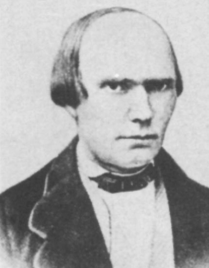
HistoryNamed after Heinrich Adolf Rinne (1819-1868), a German otologist who received his medical doctorate from the University of Göttingen. He would practice here for the majority of his career exploring the diseases of the ears, nose, and throat. He first described his eponymous test in 1855, but did not get widespread recognition for it until 1881 when it was further publicized by otologists Bezold and Lucae


References

  1. Firkin BG and Whitwirth JA.  Dictionary of Medical Eponyms. 2nd ed.  New York, NY; Parthenon Publishing Group. 1996.
  2. Bartolucci S, Forbis P.  Stedman’s Medical Eponyms.  2nd ed.  Baltimore, MD; LWW.  2005.
  3. Yee AJ, Pfiffner P. (2012).  Medical Eponyms (Version 1.4.2) [Mobile Application Software].  Retrieved http://itunes.apple.com.
  4. Whonamedit – dictionary of medical eponyms. http://www.whonamedit.com
  5. Up To Date. www.uptodate.com
  6. Heck WE. Dr. A. Rinne. Laryngoscope. 1962;72(5):647-652. [link]

#67 – Epistaxis


***LISTEN TO THE PODCAST HERE***


Anatomy

Anterior

  • Kiesselbach’s Plexus (Little’s area)
    • Confluence of three main vessels
      • Septal branch of the anterior ethmoidal artery
      • Lateral nasal branch of the sphenopalatine artery
      • Septal branch of the superior labial branch of the facial artery

Posterior

  • Woodruff’s Plexus
    • Posteriorlateral branches of the sphenopalatine artery
      • Posterior inferior turbinate

Epidemiology

  • Up to 60% of population will experience a significant nosebleed each year
    • Only 10% need to seek attention
  • Common ENT admission condition, but rarely needs surgical intervention
  • Bimodal age distribution
    • Before 10 years or between 45-65 years
  • Male predominance before the age of 49, then equalizes
    • Estrogen has been shown to protective for mucosa
  • Anterior bleeds are significantly more common (>90%) and resolve with minor interventions
  • Posterior bleeds can result in significant hemorrhage

Etiologies

  • Nose picking
  • Low environmental moisture
  • Mucosal hyperemia of viral or allergic rhinitis
  • Trauma
  • Foreign body
  • Anticoagulation
  • Coagulopathies
    • Osler-Weber-Rendu, von Willebrand, hemophilias
  • Connective tissue disease
    • Aneurysm development
  • Neoplasm
    • Squamous cell, inverted papilloma
  • Hypertension
    • Debated as a cause, but has shown to prolong bleeding
  • Nasal medications
    • Steroids, oxymetazoline
  • Heart failure

Patient Assessment

  • Primary
    • Airway assessment
      • RR, O2
    • Cardiovascular stability
      • HR, BP
  • Secondary
    • History
      • Medications
        • Anticoagulation, aspirin, nasal medications
      • PMH
        • Bleeding disorders, HTN, liver disease
        • Recent trauma
        • History of nosebleeds
          • How often, how long do they last, ever been admitted for one
  • Diagnostic Studies
    • Coagulation studies should NOT be routinely ordered
      • Should be in patients on anticoagulation
    • In patients with prolonged bleeds:
      • CBC
      • Type and cross
  • Examination
    • Have patient blow nose to remove clots and blood
    • Examine nasal cavity to see if you can see the bleeding site
      • Otoscope, nasal speculum
      • Don’t have patient tilt head back
        • Nasopharynx lies in anteroposterior plane and this will obscure the majority of the cavity from view

Interventions

  • Initial (Woodpecker/Walrus technique)
    • Have patient blow nose to remove clots
    • In a small basin mix any or all of the following:
      • Oxymetazoline
      • Lidocaine with epinephrine
      • Tranexamic acid
      • If available, soak GelFoam/Surgicel in this fluid and place BEFORE the sponge sticks
    • Trim two oral sponge swabs to better fit in the nasal cavity and soak in the fluid
    • Make a nasal bridge clamp by taping two tongue depressors together on one end
    • Place swabs in nasal cavities and apply nasal clamp for 10-15 minutes
    • Ice pack can also be used
  • Cautery
    • If the bleeding site can be visualized on direct examination
    • Apply topical anesthetic
    • Silver nitrate sticks
      • Start from periphery and roll to center of bleeding
      • No more than 10 seconds
      • A white eschar should form
  • Nasal packing
    • Use if cautery fails
    • Ensure topical anesthesia
    • Soak in sterile water
    • Insert by sliding along the floor of the nasal cavity PARALLEL to floor
    • Insufflate the balloon with air
  • Nasal Balloon Catheters
    • For posterior bleeds
    • Follow same steps for nasal packing
    • Insufflate posterior balloon FIRST and apply gently traction
    • Then insufflate the anterior balloon
  • Foley Catheters
    • If you don’t have a prefabricated nasal balloons, a foley catheter can work
    • Insert the catheter until you can see it in the posterior oropharynx
    • Insufflate with 5-10cc of water
    • Apply traction to seat balloon in posterior choana
    • Add additional water to tamponade
    • Clamp catheter with umbilical clamp or c-clamp from NG tube

Disposition and Follow-up

  • For simple nasal packing, patients should be evaluated by ENT within 24-48 hours
    • Discuss with consultant need for antibiotic prophylaxis
      • No good evidence supports routine use, but ENT often prefers
        • Amoxicillin-Clavulanate is most commonly used
        • Clindamycin or trimethoprim/sulfamethoxazole should be used if concern for nasal carrier of MRSA
  • Posterior bleeds should be immediately assessed by ENT for potential surgical intervention
    • Endoscopic sphenopalatine artery ligation
    • Anterior ethmoid artery ligation
      • Open or endoscopic

1893 Cottage Physician

References

  1. Kucik CJ, Clenney T. Management of epistaxis. Am Fam Physician. 2005; 71(2):305-11. [pubmed]
  2. Villwock JA, Jones K. Recent Trends in Epistaxis Management in the United States JAMA Otolaryngol Head Neck Surg. 2013; 139(12):1279-84. [pubmed]
  3. Kotecha B, Fowler S, Harkness P, Walmsley J, Brown P, Topham J. Management of epistaxis: a national survey. Ann R Coll Surg Engl. 1996; 78(5):444-6. [PDF]
  4. Fishpool SJ, Tomkinson A. Patterns of hospital admission with epistaxis for 26,725 patients over an 18-year period in Wales, UK. Ann R Coll Surg Engl. 2012; 94(8):559-62. [PDF]
  5. Min HJ, Kang H, Choi GJ, Kim KS. Association between Hypertension and Epistaxis: Systematic Review and Meta-analysis. Otolaryngol Head Neck Surg. 2017; 157(6):921-927. [pubmed]
  6. Shakeel M, Trinidade A, Iddamalgoda T, Supriya M, Ah-See KW. Routine clotting screen has no role in the management of epistaxis: reiterating the point. Eur Arch Otorhinolaryngol. 2010; 267(10):1641-4. [pubmed]
  7. Lin G, Bleier B. Surgical Management of Severe Epistaxis. Otolaryngol Clin North Am. 2016; 49(3):627-37. [pubmed]

PAINE #PANCE Pearl – HEENT


Question

62yo man, with a history of pseudotumor cerebri, presents to your clinic with progressive headache and vision changes. You would like to confirm an increased intracranial pressure before sending him to the neurologist.

  1. What are two (2) ways at the bedside you can confirm and what are the thresholds for positive findings?

#66 – How to be a Good Student on Emergency Medicine Rotation


***LISTEN TO THE PODCAST HERE***


Guest for the Episode

John B. Hurt, MPAS, PA-C

Assistant Professor and Director of Academics and Curriculum – Samford University – Department of Physician Assistant Studies


My 10 Rules

  1. Be on time
    • On-time = early
  2. Introduce yourself to the team
    • Preceptor and/or supervising physician
    • Charge nurse
    • Secretary or unit clerk
  3. Be Goal Oriented
    • Shift
      • Have 1-2 objectives for every shift you work
    • Rotation
      • Talk with your preceptor about what you want to see/do/experience
        • Actively seek out these experience
  4. Always Be Doing Something
    • Checking labs/images
    • Ask to draw blood for techs
    • Ask if you can get anything for your preceptor/staff
    • Review cases
      • There is something to be learned about every case we see in the ED
      • Look them up, write them down
      • Ask your preceptor what their decision making looks like
  5. Develop an algorithmic approach to common ED presentations
    • Chest pain, abdominal pain, AMS, fever, dyspnea, back pain, nausea/vomiting, trauma
      • Fast the providers what they do
      • Write these out to reference in the future
      • Get familiar with clinical decision instruments
        • Ottawa Rules, NEXUS, PERC, Canadian C-Spine
  6. 1-minute Presentations are an absolutely must
    • Have your diagnosis and plan ready
    • Differential diagnosis
      • Life threats, most likely, plausible, Zebras
  7. Disposition is the end decision in EM
    • Every patient either gets admitted or discharged
    • Before your preceptor makes the decision, make your own and see if your right
  8. See every type of patient that walks through the door
    • Don’t cherry pick (unless it is on your bucket list)
    • This will help you immensely throughout your career
  9. Go up and see the patients you admit before you leave the hospital after a shift
    • It will benefit your decision in the long run if you see what the inpatient team is doing
    • Your patients will also appreciate you checking on them
  10. Get feedback after every shift (if possible)
    • Plus-Delta approach
      • Things you do well (plus)
      • Things you can change (delta)


Ep-PAINE-nym


Puestow Procedure

Other Known Aliases – Puestow-Gillesby procedure, lateral pancreaticojejunostomy

Definitionside-to-side anastomosis of the main pancreatic duct of Wirsung to the proximal jejunum

Clinical Significance this is a surgical management option for patients with chronic pancreatitis by simultaneously facilitating drainage and preserving physiologic function of the pancreas.

HistoryNamed after Charles Bernard Puestow (1902-1973), an American surgeon who recieved his medical doctorate from the University of Pennsylvania in 1925. He would serve as a military surgeon during the 2nd World War and commanded the 27th Evacuation Hospital providing surgical services to wounded soldiers in Europe and North Africa. His commitment to the veteran population would continue after the war when he established the first surgical residency program based in a veterans hospitals in the United States in 1946. It was at Hines Veterans Hospital in Illinois where he and his partner, William Gillesby, would publish their experience and outcomes on 21 patients with chronic pancreatitis in 1958, which would lead to the creation of his eponymonic surgical procedure.


References

  1. Firkin BG and Whitwirth JA.  Dictionary of Medical Eponyms. 2nd ed.  New York, NY; Parthenon Publishing Group. 1996.
  2. Bartolucci S, Forbis P.  Stedman’s Medical Eponyms.  2nd ed.  Baltimore, MD; LWW.  2005.
  3. Yee AJ, Pfiffner P. (2012).  Medical Eponyms (Version 1.4.2) [Mobile Application Software].  Retrieved http://itunes.apple.com.
  4. Whonamedit – dictionary of medical eponyms. http://www.whonamedit.com
  5. Up To Date. www.uptodate.com
  6. Bosmia AN, Christein JD. Charles Bernard Puestow (1902-1973): American surgeon and commander of the 27th Evacuation Hospital during the Second World War. J Med Biogr. 2017; 25(3):147-152. [pubmed]
  7. PUESTOW CB, GILLESBY WJ. Retrograde surgical drainage of pancreas for chronic relapsing pancreatitis. AMA Arch Surg. 1958; 76(6):898-907. [pubmed]

PAINE #PANCE Pearl – Gastrointestinal


Question

We all know that nutritional supplementation during pregnancy is extremely important for fetal development. But…….there is one particular vitamin that can be teratogenic in excess doses.

  1. What vitamin is this?
  2. What types of foods are extremely high in this vitamin and why?


Question

  1. The class of retinoic acids have two main forms of vitamin A:
    1. Provitamin A – primarily plant based carotenoids
      1. green leafy vegetables, sweet potatoes, carrots
    2. Preformed vitamin A – primarily in animal sources
      1. livers, kidneys
  2. Up to 85% of metabolized vitamin A is stored in the liver and other tissues due to being fat soluble. As a result, animal organs (liver, kidneys, thymus) are extremely high in preformed vitamin A and can cause acute and/or chronic toxicity when ingested in large quantities.

References

  1. Rothman KJ, Moore LL, Singer MR, Nguyen UD, Mannino S, Milunsky A. Teratogenicity of High Vitamin A Intake N Engl J Med. 1995; 333(21):1369-1373.

Ep-PAINE-nym


Ranson’s Criteria

Other Known Aliases – none

Definitionclinical decision instrument to predict mortality of acute pancreatitis on admission and after the first 48 hours

Clinical Significance this was one of the first instruments to help with the initial management of patients with acute pancreatitis. Now, it has been largely been replaced by more accurate and reliable calculations and is taught only for historical purposes.

History – Named after John H. C. Ranson (1938-1995), an English-American surgeon who received his medical doctorate from Oxford University in 1960. He would complete his surgical residency at Bellevue Hospital and New York University Medical Center, where he would join as faculty and later as the Director of the Division of General Surgery. He would have a prolific career primarily focusing on the alimentary tract with concentration on the pancreas. He would publish his eponymous scoring system in 1974 which not only improved the clinical care of patients with pancreatitis, but also improved the quality of the research by finally being able to compare severity groups of treatment arms.


References

  1. Firkin BG and Whitwirth JA.  Dictionary of Medical Eponyms. 2nd ed.  New York, NY; Parthenon Publishing Group. 1996.
  2. Bartolucci S, Forbis P.  Stedman’s Medical Eponyms.  2nd ed.  Baltimore, MD; LWW.  2005.
  3. Yee AJ, Pfiffner P. (2012).  Medical Eponyms (Version 1.4.2) [Mobile Application Software].  Retrieved http://itunes.apple.com.
  4. Whonamedit – dictionary of medical eponyms. http://www.whonamedit.com
  5. Up To Date. www.uptodate.com
  6. Reber HA. Obituary – John H. C. Ranson, Pancreas: April 1996 – Volume 12 – Issue 3 – p 215 [link]
  7. Ranson JH, Rifkind KM, Roses DF, Fink SD, Eng K, Spencer FC. Prognostic signs and the role of operative management in acute pancreatitis. Surg Gynecol Obstet. 1974;139(1):69-81. [link]

Ep-PAINE-nym


Barrett’s Esophagus

Other Known Aliases – Allison-Johnstone anomaly

Definitionmetaplastic change of the mucosal cells of the lower esophagus from normal stratified squamous epithelium to simple colunar epithelium and interspaced goblet cells

Clinical Significance these histologic changes are premaligant and significantly increases a patient’s risk for developing esophageal adenocarcinoma.

HistoryNamed after Norman Rupert Barrett (1903-1979), an Australian-born British thoracic surgeon who received his medical doctorate from Trinity College, Cambridge. He would practice his entire career at St. Thomas Hospital, with a brief training period in 1935-1936 when he traveled to the US on a Rockfeller Traveling Fellowship. It was here that he decided to pursue thoracic surgery instead of GI surgery. In 1947, he performed the first successful surgical repair of a ruptured esophagus. He would publish his eponymous findings of histologic changes of the distal esophagus in 1950, but erroneously believed this was due to congenitally shortened esophagus with a portion of the stomach trapped in the chest. Allison and Johnstone were the first to argue that these changes were esophagus, not stomach, and termed these ulcers “Barrett’s ulcers”. Of note, Allison first described this condition in 1948 before Barrett’s publication.


References

  1. Firkin BG and Whitwirth JA.  Dictionary of Medical Eponyms. 2nd ed.  New York, NY; Parthenon Publishing Group. 1996.
  2. Bartolucci S, Forbis P.  Stedman’s Medical Eponyms.  2nd ed.  Baltimore, MD; LWW.  2005.
  3. Yee AJ, Pfiffner P. (2012).  Medical Eponyms (Version 1.4.2) [Mobile Application Software].  Retrieved http://itunes.apple.com.
  4. Whonamedit – dictionary of medical eponyms. http://www.whonamedit.com
  5. Up To Date. www.uptodate.com
  6. Barrett NR. Chronic peptic ulcer of the oesophagus and oesophagitis. British Journal of Surgery. 1950;38:175-182. [link]
  7. P. R. Allison, A. S. Johnstone. The esophagus lined with gastric mucous membrane. Thorax, 1953, 8: 87.
  8. P. R. Allison. Peptic ulcer of the Oesopahgus. Thorax, 1948, 3: 20.

#65 – Pancreatitis


***LISTEN TO THE PODCAST HERE***


Epidemiology

  • 13-45 per 100,000 person incidence in the US
  • Most common GI cause of hospital admission in the US
    • > 300,000 per year
    • Average 4-day stay with cost > $6000/case
  • Equal gender representation across the lifespan
    • Alcohol pancreatitis more common in men
  • 2-3 fold higher rates in African Americans

Risk Factors and Etiologies

  • Gallstones
    • 40-70% of cases
      • Only 3-7% of patients with gallstones develop pancreatitis
    • Two theorized mechanisms
      • Reflux of bile into the pancreatic duct
      • Obstruction at the ampulla
    • Magic number is 5mm
      • Small enough to pass through cystic duct, but still get obstructed at ampulla
  • Alcohol
    • 25-35% of cases
    • Interesting data to show that it is not just alcohol that causes pancreatitis
      • 5 out of 100,00 patents with alcohol abuse develop pancreatitis
    • Several mechanisms theorized
      • Sensitization of acinar cell to CCK-induced activation of zymogens
      • Potentiation of the effect of CCK
      • Generation of toxic metabolites
      • Sensitization of the pancreas to toxic insults
      • Activation of pancreatic stellate cells to increase production of matrix proteins
  • Idiopathic (genetic)
    • 15-25% of patients with pancreatitis have no identifiable pathologic cause
    • These cases are largely theorized to have complex genetic risk profiles
  • Hypertriglyceridemia
    • 1-14% of cases
    • > 1000 mg/dL increases risk
  • Post-ERCP
    • 3% of patients undergoing diagnostic ERCP
    • 5% of patients undergoing therapeutic ERCP
    • 25% of patients undergoing sphincter of Oddi measurements
  • Medications
    • < 5% of cases
    • Classification system (Ia, Ib, II, III, IV)
    • Prognosis is excellent and mortality is very low
  • Obesity
  • Smoking
  • Diabetes

Pathogenesis

  • Pancreatic enzymes synthesis continues while secretion is slowed or halted
  • HIT #1 –  Intraacinar activation of proteolytic enzymes (trypsin)
    • Cascade of enzyme release and activation then occurs
    • Ultimately, causes autodigestion of the pancreas
  • HIT #2 – Microcirculatory injury
    • Damage to the pancreas via autodigestion leads to vasoconstriction, decreased oxygenation, and progressive ischemia
      • Leads to edema and further decreased secretion of enzymes
  • HIT #3 – Leukocyte infiltration, cytokine release, and oxidative stress
    • Leads to widespread inflammation and induce thrombosis and hemorrhage
    • Ultimately, causes necrosis
  • Two main classifications
    • Interstitial pancreatitis – blood supply is maintained
    • Necrotizing pancreatitis – blood supply is affected

Signs and Symptoms

  • History
    • Epigastric pain
      • May radiate to the back
      • May radiate to the right shoulder
        • Kehr’s sign
    • Nausea
    • Vomiting
    • Dyspneic
      • Severe disease can cause diaphragmatic irritation and pleural effusions
  • Physical Examination
    • Fever
    • Tachycardia
    • Epigastric tenderness
    • Abdominal distention
    • Hypoactive bowel sounds
    • Jaundiced
    • Abdominal ecchymosis (necrotizing disease)
      • Cullen’ sign – umbilical
      • Grey Turner’s sign – flank
      • Fox’s sign – thigh (parallel but inferior to inguinal ligament)

Laboratory Studies

  • Serum amylase
    • Rises within 6 hours, returns to normal in 3-5 days
    • Should not be used (sensitivity 67-83%, specificity 85-98%)
      • Short-half life (10 hours)
        • Patients presenting > 24 hours after onset can have normal amylase
      • Miss up to 20% of cases of alcohol pancreatitis
        • Due to inability of parenchyma to produce amylase
      • Miss up to 50% of cases of hypertriglyceridemia
        • Triglycerides interfere with assay
  • Serum lipase
    • Sensitivity 85-100%
    • Rises 4-8 hours, peaks at 24 hours, returns to normal in 8-14 days
  • LFTs
    • Evaluate for cholestatic elevations (ALP, bilirubin, GGT)
  • BMP
    • Need glucose, BUN, and calcium for some of the risk calculators
  • CBC
    • Leukocytosis often is present and helps grade severity
    • May show hemoconcentration due to volume depletion

Imaging Studies

  • Ultrasound
    • Often the quickest and easiest study to obtain in the ED
    • Can evaluate gallbladder pathology, stones, and peripancreatic fluid
    • Ileus can obscure imaging due to gas overlying the pancreas
  • CT
    • Better detail and can evaluate more structures
  • MRI
    • Higher sensitivity in early disease
    • Longer to obtain
  • Revised Atlanta Criteria
    • Six CT morphological features
    • Interstitial edema
      • Parenchymal enhancement by IV contrast
    • Necrotizing findings
      • Lack of parenchymal enhancement
      • Peripancreatic fluid collection or walled-off necrosis
    • Acute peripancreatic fluid collection
      • Homogenous fluid collection
      • Confined to normal peripancreatic fascial planes
      • No definable encapsulating wall
      • Adjacent to pancreas (no intrapancreatic extension)
    • Pancreatic pseudocyst
      • Well-circumscribed, well-defined wall with homogenous fluid density
      • No non-liquid component
    • Acute necrotic collection
      • Heterogenous with non-liquid density of varying degrees
      • No definable wall
      • Intra-, or extra-pancreatic in location
    • Walled-off necrosis
      • Heterogenous with liquid and non-liquid densities
      • Well-defined wall that is completely encapsulated
      • Intra-, or extra-pancreatic in location

Diagnosis

  • Need 2 of the following 3 criteria:
    • Acute onset of persistent, severe, epigastric pain
    • Elevation of amylase or lipase > 3x ULN
    • Radiographic findings on imaging

Classification of Severity

  • Mild
    • Absence of organ failure or local/systemic complications
  • Moderately severe
    • Transient organ failure (resolves with 48 hours) and/or systemic complications without persistent organ failure
  • Severe
    • Persistent organ failure

Prognosis Predictor Scoring Systems

  • Ranson’s Criteria
  • BISAP Score
    • 0-2 points – low mortality
    • 3-5 points – high mortality

Management

  • Patients with mild pancreatitis can be admitted to floor/wards
  • Patients with moderately severe or severe should be admitted to ICU
  • Fluid resuscitation
    • 5-10 mL/kg/hour with crystalloid
      • Careful, using LR in patients with hypercalcemic induced pancreatitis
    • Bolus 20 ml/kg over 30 minutes if hypotensive/tachycardic
    • Adjust using goal-directed metrics even 6 hours for first 24 hours-48 hours
      • BUN, H/H, MAP (65-85 mmHg), HR (< 120bpm), UOP (>0.5 mL/kg/hr)
  • Pain control
    • Opioids are safe and PCA can work well
      • Fentanyl has better safety profile
        • 20-50 mcg with 10-min lockout
  • Nutrition
    • Can reintroduce within 24 hours if no nausea, vomiting, and decreasing pain and inflammatory markers
      • Start with low-residue, low fat, soft diet and advance as tolerated
    • Supplemental nutrition generally needed for moderately severe and severe cases, or if unable to tolerate oral nutrition within 5 days
      • Enteral > parental with placement of jejunal feeding tube beyond the ligament of Treitz
        • Helps prevent bacterial translocation
      • Parenteral is indicated if nutritional goals are not achieved with 48-72 hours due to pain or intolerance
      • Consult your hospital nutritional team and/or dietician for help
  • Antibiotics
    • No evidence to support prophylactic antibiotics
      • Most infected necroses will occur late in clinical course (5-10 days after admission)
  • Treat underlying causes
    • Gallstone pancreatitis
      • ERCP should be performed within 24 hours of admission
      • Cholecystectomy should be performed within 7 days and often during same hospitalization
    • Hypertriglyceridemia
      • Therapeutic plasma exchange and insulin therapy

Complications

  • Necrosis
  • Pseudocyst
  • Splanchnic venous thrombosis


References

  1. Yadav D, Lowenfels AB. The epidemiology of pancreatitis and pancreatic cancer. Gastroenterology. 2013; 144(6):1252-61. [PDF]
  2. Conwell DL, Banks PA, Greenberger NJ. Acute and Chronic Pancreatitis. In: Jameson J, Fauci AS, Kasper DL, Hauser SL, Longo DL, Loscalzo J. eds. Harrison’s Principles of Internal Medicine, 20e. McGraw-Hill;
  3. Mechanisms of alcoholic pancreatitis. Proceedings of a conference. Chicago, Illinois, USA, November 2002. Pancreas. 2003; 27(4):281-355. [pubmed]
  4. Nawaz H, Koutroumpakis E, Easler J, et al. Elevated serum triglycerides are independently associated with persistent organ failure in acute pancreatitis. Am J Gastroenterol. 2015; 110(10):1497-503. [pubmed]
  5. Scherer J, Singh VP, Pitchumoni CS, Yadav D. Issues in hypertriglyceridemic pancreatitis: an update. J Clin Gastroenterol. 2014; 48(3):195-203. [PDF]
  6. Kahaleh M, Freeman M. Prevention and management of post-endoscopic retrograde cholangiopancreatography complications. Clin Endosc. 2012; 45(3):305-12. [PDF]
  7. Lankisch PG, Dröge M, Gottesleben F. Drug induced acute pancreatitis: incidence and severity. Gut. 1995; 37(4):565-7. [PDF]
  8. Forsmark CE, Swaroop Vege S, Wilcox CM. Acute Pancreatitis N Engl J Med. 2016; 375(20):1972-1981.
  9. Yadav D, Agarwal N, Pitchumoni CS. A critical evaluation of laboratory tests in acute pancreatitis. Am J Gastroenterol. 2002; 97(6):1309-18. [pubmed]
  10. Wu BU, Johannes RS, Sun X, Tabak Y, Conwell DL, Banks PA. The early prediction of mortality in acute pancreatitis: a large population-based study. Gut. 2008; 57(12):1698-703. [pubmed]
  11. Vege SS, DiMagno MJ, Forsmark CE, Martel M, Barkun AN. Initial Medical Treatment of Acute Pancreatitis: American Gastroenterological Association Institute Technical Review. Gastroenterology. 2018; 154(4):1103-1139. [pubmed]
  12. Basurto Ona X, Rigau Comas D, Urrútia G. Opioids for acute pancreatitis pain. Cochrane Database Syst Rev. 2013; [pubmed]
  13. Casaer MP, Mesotten D, Hermans G, et al. Early versus late parenteral nutrition in critically ill adults. N Engl J Med. 2011; 365(6):506-17. [pubmed]
  14. Kutsogiannis J, Alberda C, Gramlich L, et al. Early use of supplemental parenteral nutrition in critically ill patients: results of an international multicenter observational study. Crit Care Med. 2011; 39(12):2691-9. [pubmed]
  15. Aboulian A, Chan T, Yaghoubian A, et al. Early cholecystectomy safely decreases hospital stay in patients with mild gallstone pancreatitis: a randomized prospective study. Ann Surg. 2010; 251(4):615-9. [pubmed]
  16. Uhl W, Müller CA, Krähenbühl L, Schmid SW, Schölzel S, Büchler MW. Acute gallstone pancreatitis: timing of laparoscopic cholecystectomy in mild and severe disease. Surg Endosc. 1999; 13(11):1070-6. [pubmed]
  17. Ipe TS, Pham HP, Williams LA 3rd. Critical updates in the 7 edition of the American Society for Apheresis guidelines. J Clin Apher. 2018; 33(1):78-94. [pubmed]